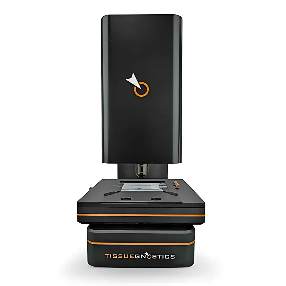
StrataFAXS II Tissue and Cell Quantitative Analysis System by TissueGnostics
| Brand | TissueGnostics |
|---|---|
| Origin | Austria |
| Manufacturer Type | Original Equipment Manufacturer (OEM) |
| Country of Origin | Imported |
| Model | StrataFAXS II |
| Price Range | USD 135,000 – 205,000 |
Overview
The StrataFAXS II Tissue and Cell Quantitative Analysis System is a high-precision, upright brightfield whole-slide imaging and quantitative pathology platform engineered for robust, reproducible single-cell analysis in formalin-fixed paraffin-embedded (FFPE) tissue sections, cytospins, cell smears, and adherent cell cultures. Built upon a modular optical architecture with motorized Z-stack acquisition and real-time tile stitching, the system implements advanced computational pathology workflows grounded in pixel-level segmentation, multi-layer morphometric feature extraction, and spatially resolved biomarker quantification. Its core measurement principle combines high-fidelity brightfield microscopy (20×–60× objectives) with deterministic image registration, enabling accurate in situ cell detection, subcellular compartment delineation (nucleus/cytoplasm/membrane), and context-aware intensity calibration aligned to standardized IHC staining protocols (e.g., DAB, AEC, hematoxylin counterstain). Designed for routine diagnostic labs and translational research facilities, StrataFAXS II delivers GLP-compliant data provenance through audit-trail-enabled acquisition logs and parameter-locked analysis pipelines.
Key Features
- Upright brightfield scanning platform with dual-slide carrier accommodating standard 1″ × 3″ glass slides or extended-format 2× slides
- Real-time on-the-fly scanning and mosaic stitching—no post-acquisition tiling delay; live preview synchronized with acquisition
- Motorized high-NA objective turret supporting 20×, 40×, and 60× dry objectives optimized for IHC, CISH, and RNAscope applications
- Integrated Z-stack focus control enabling depth-aware nuclear segmentation across variable tissue thicknesses (up to 10 µm section depth)
- StrataQuest Base software framework with role-based user authentication, encrypted project folders, and CFR Part 11–compliant audit trails for parameter changes and report generation
- One-click workflow activation via flat-panel App launcher—each APP encapsulates a validated, version-controlled analysis protocol (e.g., IHC 2 APP, IHC 3 APP, IHC Membrane APP)
Sample Compatibility & Compliance
The StrataFAXS II supports broad sample compatibility including FFPE tissue sections (3–10 µm), frozen sections, cytology preparations (Pap smears, bronchial washings), and monolayer cell cultures grown on standard microscope slides or chambered coverslips. All APPs are developed and internally validated per ISO/IEC 17025 principles and align with analytical requirements outlined in CAP Checklist ANP.42300 (quantitative IHC), USP (image-based assay validation), and CLSI EP17-A2 (limit of detection for low-abundance markers). System firmware and StrataQuest Base v5.3+ support FDA 21 CFR Part 11 electronic signatures, secure user access tiers (Admin / Analyst / Viewer), and immutable metadata embedding into exported PDF reports (including acquisition timestamps, objective ID, exposure settings, and APP version hash).
Software & Data Management
StrataQuest Base serves as the unified analytical engine, integrating image acquisition, batch processing, and statistical reporting within a single GUI. Each APP operates as an isolated computational module—parameters are preconfigured, locked during execution, and stored with cryptographic integrity. The two-dimensional flow-like scatter plot interface enables dynamic gating (Gate/Input Gate) with bidirectional image–data linkage: selecting a cluster updates the viewport to highlight corresponding cells in the original slide; conversely, clicking any cell in the image instantly highlights its coordinates in the scatter plot. All extracted features—including nuclear area, cytoplasmic DAB optical density, membrane circumference, aspect ratio, and integrated intensity—are stored in structured SQLite databases. Batch export supports CSV (for R/Python integration), annotated TIFF (with overlay masks), and regulatory-grade PDF reports containing raw metrics, histogram distributions, spatial heatmaps, and QC flags.
Applications
- Quantitative immunohistochemistry: automated enumeration of PD-L1+, CD8+, Ki-67+, ER/PR+ cell populations with spatial density mapping across tumor/stroma compartments
- Chromogenic in situ hybridization (CISH) and RNAscope signal dot counting—including subnuclear localization scoring and co-expression analysis in multiplexed assays
- Morphometric profiling of epithelial-mesenchymal transition (EMT) markers via simultaneous nuclear shape index and membrane E-cadherin intensity quantification
- Preclinical toxicology studies requiring longitudinal cell-counting consistency across multi-site histopathology cores
- Clinical trial biomarker assay development where SOP-driven repeatability and inter-laboratory concordance are mandated
FAQ
Does StrataFAXS II support fluorescence imaging?
No—StrataFAXS II is optimized exclusively for brightfield modalities (IHC, H&E, trichrome, CISH). Fluorescence capabilities require the complementary StrataFLUX platform.
Can third-party algorithms be imported into StrataQuest Base?
Yes—via the StrataSDK developer toolkit, which provides C++/Python APIs for custom feature extraction modules compliant with StrataQuest’s plugin architecture and metadata schema.
Is remote system monitoring and control supported?
Yes—through TLS-secured StrataLink web interface, enabling real-time status dashboards, queue management, and supervised remote analysis session handover without compromising local data residency.
How is calibration traceability maintained across instruments?
Each system ships with NIST-traceable color and focus calibration slides; StrataCalibrate utility performs daily auto-validation of white balance, intensity linearity, and Z-step accuracy—with logs archived alongside project data.
What file formats are natively supported for import/export?
Native acquisition format is .tgs (TissueGnostics Slide); imports include SVS, NDPI, MRXS, TIFF (with OME-TIFF metadata); exports include PDF, CSV, TIFF (8/16-bit), and JSON-structured feature sets compatible with QIAGEN CLC Biomedical Workbench and HALO® interoperability plugins.


