Tucsen Dhyana 400D Scientific CMOS Camera for Fluorescence Imaging of Bone, Renal, and Neural Cells
| Brand | Tucsen |
|---|---|
| Origin | Fujian, China |
| Manufacturer Type | Original Equipment Manufacturer (OEM) |
| Origin Category | Domestic |
| Model | Dhyana 400D |
| Pricing | Upon Request |
| Image Resolution | 2048 × 2048 |
| Pixel Size | 6.5 µm × 6.5 µm |
| Sensor Format | 1.2″ |
| Readout Speed | 35 fps |
| Dynamic Range | 85 dB |
Overview
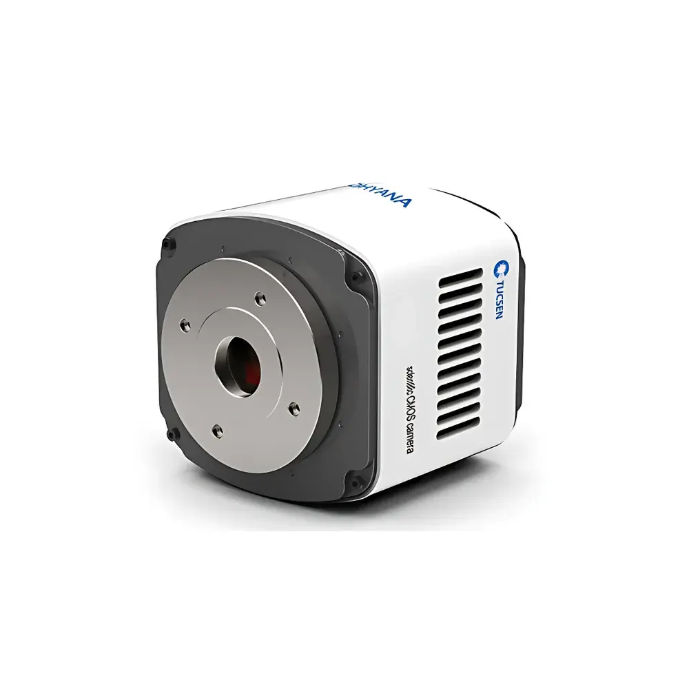
The Tucsen Dhyana 400D is a high-performance scientific CMOS camera engineered for quantitative fluorescence microscopy in life science research laboratories. Built around the G2020e sCMOS sensor—a 1.2″ format, monochrome imaging device—the Dhyana 400D delivers exceptional photon capture efficiency with a peak quantum efficiency of 72% at 595 nm and up to 80% across key visible and near-visible emission bands. Its 2048 × 2048 active pixel array, coupled with 6.5 µm square pixels and a 13.3 mm × 13.3 mm photosensitive area, provides an optimal balance between field-of-view coverage and spatial sampling resolution when paired with standard C-mount microscope adapters. The camera employs a rolling shutter architecture with hardware-accelerated 2×2 pixel binning—implemented at the analog front-end—to enhance signal-to-noise ratio (SNR) without sacrificing frame integrity or introducing interpolation artifacts. This capability makes it particularly suitable for low-light applications such as time-lapse imaging of live bone-derived mesenchymal stromal cells, calcium dynamics in primary renal tubular epithelial cells, and sparse neuronal activity mapping in brain slice preparations.
Key Features
- Monochrome sCMOS sensor (G2020e) with 1.2″ optical format and 2048 × 2048 resolution
- Pixel size: 6.5 µm × 6.5 µm; full-well capacity: 45 ke⁻ (typical)
- Read noise: 2 e⁻ (typical, 16-bit mode); dark current: 0.12 e⁻/pixel/s at –10°C
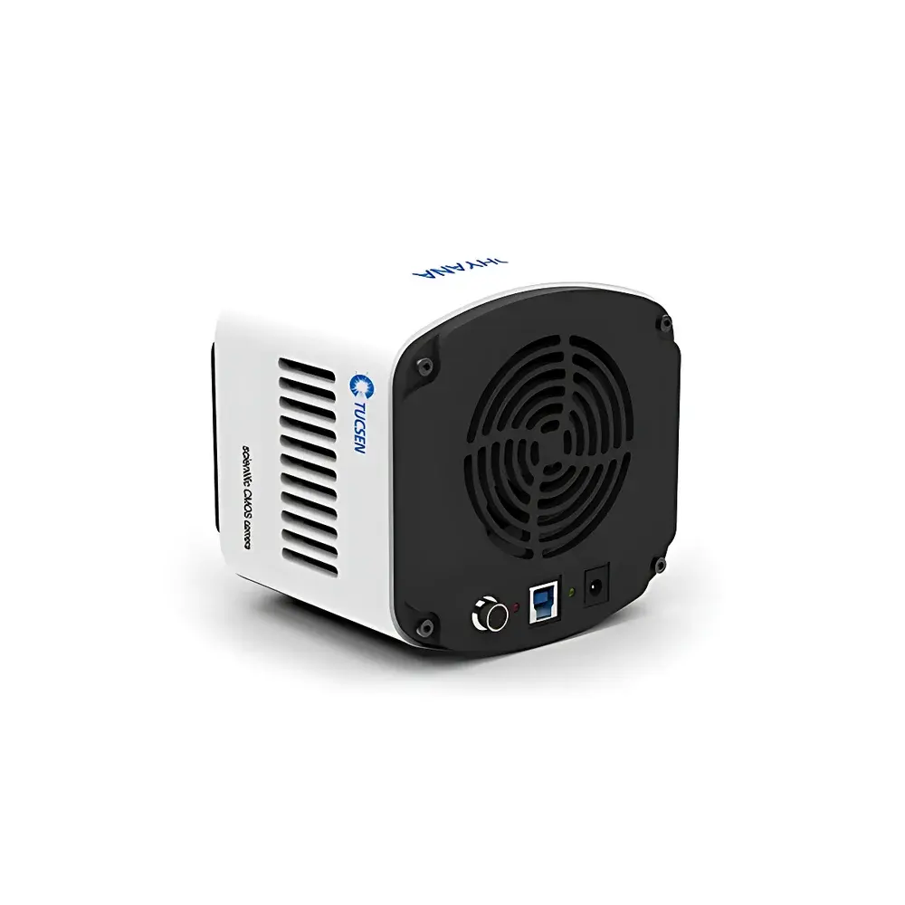
- Air-cooled thermal management: stabilizes sensor temperature up to 35°C below ambient
- Dynamic range: 86.6 dB (calculated from full-well capacity and read noise)
- Frame rate: 35 fps at full resolution and 16-bit depth
- Hardware-based 2×2 binning for improved sensitivity in photon-starved conditions
- Flexible exposure control: 13 µs to 10 s, supporting both manual and trigger-synchronized acquisition
- Multi-mode triggering: standard, synchronous, global reset, and software-triggered exposure output via SMA interface
- C-mount lens interface compatible with standard inverted and upright fluorescence microscopes
Sample Compatibility & Compliance
The Dhyana 400D is validated for use in fixed and live-cell fluorescence imaging workflows involving adherent and suspension cultures—including osteoblasts, podocytes, proximal tubule cells, primary cortical neurons, and organotypic brain slices. Its low read noise and high dynamic range support quantitative intensity measurements required for ratiometric calcium imaging (e.g., Fura-2, GCaMP), immunofluorescence colocalization analysis, and long-term mitotic tracking. While the camera itself is not a medical device, its performance characteristics align with ISO 13660 (imaging system resolution standards) and ASTM E2912 (standard guide for evaluating digital imaging systems in biological microscopy). Data acquisition protocols using this camera may be documented to satisfy GLP-compliant recordkeeping requirements, especially when integrated with timestamped metadata logging in supported software environments.
Software & Data Management
The Dhyana 400D is supported by Tucsen’s native Mosaic acquisition suite, offering real-time preview, multi-channel time-series capture, ROI-based histogram analysis, and non-destructive bit-depth scaling. It provides native SDKs for C, C++, C#, and Python, enabling seamless integration into custom pipelines for automated image analysis, machine learning–based segmentation, or synchronization with electrophysiology rigs. Third-party compatibility includes Micro-Manager 2.0 (v2.0.2+), LabVIEW (NI Vision 2020+), and MATLAB Image Acquisition Toolbox (R2021a+). All drivers support Windows 10/11 (64-bit) and select Linux distributions (Ubuntu 20.04 LTS, CentOS 7.9). Metadata embedding follows TIFF 6.0 and OME-TIFF specifications, ensuring traceability and interoperability with open-source platforms such as QuPath and Napari.
Applications
- Live-cell fluorescence imaging of intracellular Ca²⁺, pH, ROS, and membrane potential indicators
- Immunofluorescent detection and quantification of cytoskeletal proteins (e.g., actin, tubulin) and nuclear markers (e.g., DAPI, Hoechst)
- Long-term time-lapse monitoring of osteogenic differentiation, nephrotoxicity assays, and neurodevelopmental processes
- Widefield deconvolution microscopy and structured illumination preparation (when used with appropriate filter sets and illumination sources)
- Quantitative colocalization analysis (Pearson’s coefficient, Manders’ overlap) in multi-label experiments
- Integration with automated stage systems for tiled whole-slide imaging of cultured cell monolayers
FAQ
Is the Dhyana 400D compatible with Nikon or Olympus C-mount microscope adapters?
Yes—the camera uses a standardized C-mount mechanical interface with 17.526 mm flange focal distance, ensuring mechanical and optical compatibility with all major OEM microscope manufacturers’ C-mount ports.
Does the camera support hardware triggering for synchronization with external light sources or shutters?
Yes—it offers three hardware trigger modes (Standard, Synchronous, Global Reset) and provides TTL-compatible exposure start/end signals via SMA connectors.
Can I acquire images in 12-bit mode to increase frame rate?
Yes—while the default acquisition is 16-bit for maximum dynamic range, the camera supports selectable 12-bit readout to achieve higher throughput where quantization precision is less critical.
What cooling performance can be expected in a typical lab environment (23°C ambient)?
With active air cooling engaged, the sensor stabilizes at approximately –10°C, reducing dark current by two orders of magnitude compared to uncooled operation.
Is there FDA 21 CFR Part 11 compliance support for regulated environments?
The camera itself does not provide electronic signature or audit trail features; however, when deployed within validated software environments (e.g., Micro-Manager with custom plugin logging), it may serve as part of a compliant imaging subsystem under site-specific validation protocols.


