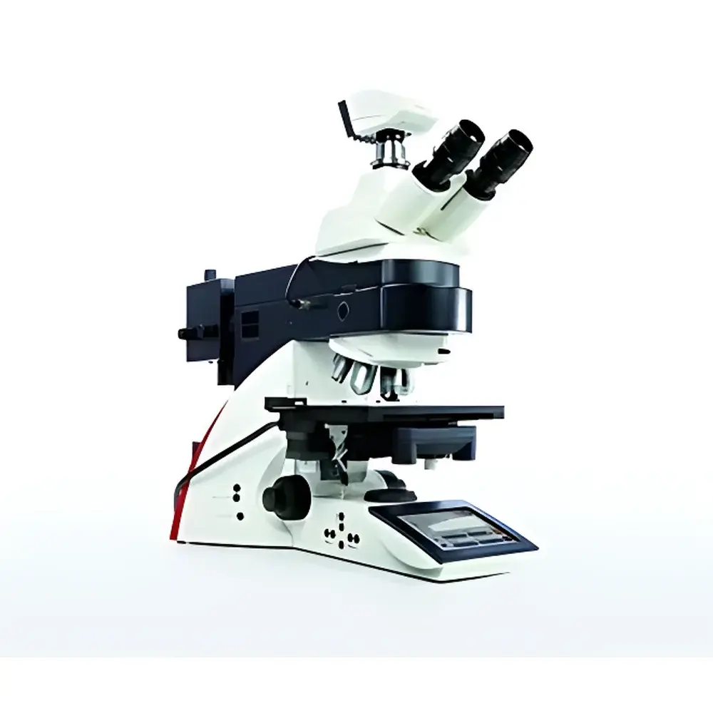
Leica DM6000B Biological Microscope
| Brand | Leica |
|---|---|
| Origin | Germany |
| Model | DM6000B |
| Optical System | Infinity-Corrected |
| Focus Drive | Motorized Z-axis with memory function |
| Objective Turret | Motorized and encoded 7-position |
| DIC Capability | Fully automated, objective-coupled DIC |
| Fluorescence | Dual excitation paths (5× or 8× filter changer), anti-bleaching intensity control |
| Illumination | 12 V / 100 W halogen transmittance light source |
| Eyepieces | 10× widefield (25 mm field number), compatible with other magnifications |
| Observation Tubes | Binocular or trinocular |
| Contrast Methods | Brightfield, Darkfield, Phase Contrast, IMC (Hoffman Modulation Contrast), Fluorescence, Differential Interference Contrast (DIC) |
| Objective Compatibility | Plan Achromat, Semi-Apochromat, Apochromat, and Long Working Distance objectives |
| Camera Interface | C-mount compatible for CCD/CMOS cameras |
| Software Integration | Compatible with LAS X, LAS AF, and third-party acquisition platforms |
| Compliance | Designed to support GLP/GMP workflows with audit-trail-capable software options (when used with LAS X Core or Advanced modules) |
Overview
The Leica DM6000B is a high-performance, research-grade biological microscope engineered for demanding life science applications requiring precision, reproducibility, and multimodal imaging capability. Built upon Leica’s proven infinity-corrected optical architecture, the DM6000B integrates motorized hardware control with intelligent optical coupling to deliver consistent, objective-matched contrast modalities—including the industry’s first fully automated Differential Interference Contrast (DIC) system. Its core design philosophy centers on eliminating manual intervention in critical alignment steps: the encoded 7-position objective turret automatically recalls optimal condenser aperture, illumination intensity, and contrast settings specific to each objective—ensuring identical optical configuration across repeated experiments and users. This level of hardware-software synchronization supports rigorous experimental protocols in cell biology, developmental studies, and live-tissue observation where quantitative repeatability and operator-independent setup are essential.
Key Features
- Motorized Z-axis with focus memory: Enables precise, repeatable focal plane positioning; stores Z-height values per objective and sample position for automated re-acquisition and Z-stack generation.
- Encoded 7-position objective turret: Synchronizes objective selection with preconfigured illumination parameters, condenser alignment, and contrast module engagement—critical for multi-contrast time-lapse or comparative studies.
- Fully automated DIC: Eliminates manual alignment of Wollaston prisms and polarizers; DIC settings are stored and recalled per objective, ensuring uniform shear direction and contrast optimization at all magnifications.
- Dual-path fluorescence illumination: Supports both 5-position and 8-position rapid filter changers with integrated intensity regulation to minimize phototoxicity and photobleaching during live-cell imaging.
- Intelligent light management: Includes color temperature stabilization for transmitted light, automatic brightness compensation across magnifications, and programmable illumination profiles for brightfield, phase, and DIC.
- Modular observation path: Trinocular port with beam-splitting ratio control allows simultaneous connection to eyepieces, digital camera, and optional confocal or spectral detectors without optical compromise.
Sample Compatibility & Compliance
The DM6000B accommodates a broad range of specimen formats—from standard glass slides and petri dishes to specialized live-cell chambers (e.g., 35 mm glass-bottom dishes) and CO2-controlled incubation stages. Its mechanical stage supports motorized XY translation with programmable coordinate mapping, enabling tile-scan acquisition over large tissue sections or multi-well plates. The platform is compatible with Leica’s environmental control systems (e.g., heating stages, humidity chambers, and gas-regulated enclosures) for extended in vivo and long-term time-lapse experiments. From a regulatory standpoint, when operated with Leica LAS X software configured in Audit Trail mode, the system supports 21 CFR Part 11-compliant data integrity requirements—including electronic signatures, user access controls, and immutable activity logs—making it suitable for quality-controlled environments adhering to GLP or GMP standards.
Software & Data Management
The DM6000B is natively supported by Leica Application Suite (LAS) X, a modular software platform designed for acquisition, analysis, and documentation in regulated and academic settings. LAS X Core provides intuitive workflow-driven control of motorized components, while LAS X Advanced adds multi-dimensional acquisition (Z-stacks, time-series, multi-channel fluorescence), spectral unmixing, and object-based quantification. All image metadata—including objective ID, exposure time, DIC bias voltage, fluorescence filter set, and focus position—is embedded directly into TIFF or LIF file headers. Export formats comply with OMERO and Bio-Formats standards, facilitating integration into institutional image repositories and AI training pipelines. Optional plugins enable direct export to MATLAB, Python (via PyLeica), and FIJI/ImageJ for custom algorithm development.
Applications
- High-content screening of fixed and live mammalian cells using multiplexed fluorescence and label-free contrast (DIC, IMC)
- Developmental biology studies requiring long-term, low-phototoxicity imaging of zebrafish, C. elegans, or organoids
- Quantitative morphometric analysis of neuronal growth cones, mitotic spindles, or organelle dynamics
- GMP-compliant QC of bioprocess-derived cell therapies, including viability, morphology, and confluence assessment
- Correlative light microscopy (CLM) workflows, serving as the optical foundation prior to EM or super-resolution validation
FAQ
Does the DM6000B support live-cell imaging under physiological conditions?
Yes—the system integrates seamlessly with motorized environmental chambers, CO2 controllers, and temperature-regulated stages. Its low-heat halogen illumination and anti-bleaching fluorescence control minimize thermal and photochemical stress.
Can DIC settings be saved and reapplied across different users or sessions?
Yes—encoded objective recognition triggers automatic recall of DIC prism orientation, bias voltage, and condenser alignment, ensuring inter-user consistency without manual recalibration.
Is the DM6000B compatible with third-party cameras and analysis software?
Yes—it features standard C-mount output and supports GenICam-compliant drivers, enabling native integration with Hamamatsu, Andor, Photometrics, and FLIR cameras, as well as Fiji, CellProfiler, and commercial AI-based analysis tools.
What software licenses are required for audit-trail functionality?
LAS X Core or Advanced with the “Audit Trail” option enabled; requires dedicated user authentication server configuration and encrypted database logging per FDA 21 CFR Part 11 guidelines.
How does the DM6000B differ from the DM6 B model?
The DM6000B offers enhanced motorization (7-position encoded turret vs. 6-position), expanded fluorescence capacity (dual-path architecture), deeper software integration (LAS X native support), and improved DIC automation fidelity compared to the earlier DM6 series.
Related Products


