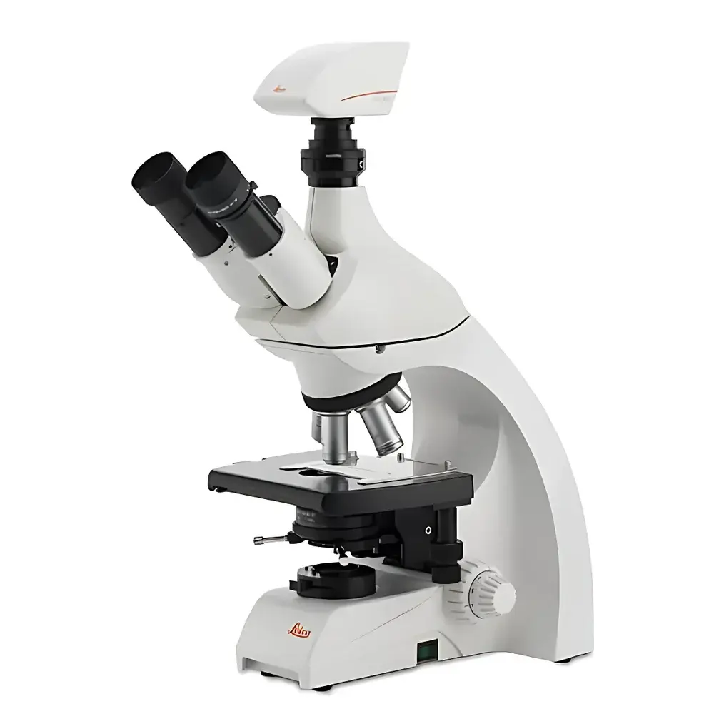
Leica DM1000 Research-Grade Biological Microscope
| Origin | Germany |
|---|---|
| Manufacturer Type | Authorized Distributor |
| Origin Category | Imported |
| Model | DM1000 |
| Pricing | Available Upon Request |
Overview
The Leica DM1000 is a research-grade biological microscope engineered for high-fidelity transmitted-light observation of fixed and live biological specimens. Built upon Leica’s HC (High Contrast) infinity-corrected optical system, it delivers exceptional image fidelity, chromatic correction, and contrast uniformity across the entire field of view. The microscope employs Köhler illumination with a 12 V / 30 W halogen lamp and integrated blue interference filter—ensuring stable, color-accurate illumination optimized for brightfield, phase contrast, and basic fluorescence applications. Its thermally compensated mechanical structure maintains optical alignment and focus stability during extended observation sessions, making it suitable for routine laboratory use in academic, clinical, and industrial life science settings.
Key Features
- HC infinity-corrected optical system with high-transmission anti-reflection coatings for enhanced resolution and contrast
- Five-position objective turret accommodating up to five standard DIN objectives
- Precision coaxial focusing mechanism with 1 µm minimum step resolution and built-in mechanical stop to prevent stage overtravel or objective collision
- Ergonomic 15° inclined trinocular head with adjustable interpupillary distance and diopter compensation on both eyepieces (10×, 22 mm field number)
- Universal Abbe condenser with adjustable aperture diaphragm and centering screws; compatible with phase contrast and darkfield accessories
- Ceramic-coated mechanical stage with gearless design, rounded corners, and single-hand specimen clip for simultaneous X/Y translation and coarse/fine focus control
- Height-adjustable focusing knobs with symmetrical layout for fatigue-free operation
- Integrated Leica DFC290 digital camera module featuring FireWire (IEEE 1394a) interface, 3073 × 2304 active pixels (7.15 MP), 3.2 × 3.2 µm pixel size, and 30-bit color depth
- Real-time imaging performance: up to 10 fps at full resolution (2048 × 1536), 25 fps at reduced resolution (1024 × 768)
Sample Compatibility & Compliance
The DM1000 supports standard glass microscope slides (1 × 3 inches / 25 × 75 mm) and coverslips (No. 1.5, 0.17 mm thickness). It is fully compatible with phase contrast techniques using Leica’s HI PLAN series objectives (4×, 10×, 20×, 40×, 100× oil, NA 0.1–1.25). All optical components comply with ISO 8578:2017 (microscope mechanical tube length and parfocality standards) and EN 61000-6-3:2019 (EMC emission requirements). The system meets general laboratory safety standards per IEC 61010-1 and is designed for integration into GLP-compliant workflows where documentation traceability and operator reproducibility are required.
Software & Data Management
The Leica DM1000 operates with Leica Application Suite (LAS) X software, supporting acquisition, annotation, measurement (length, area, intensity profiling), multi-channel overlay, time-lapse sequencing, and export in TIFF, JPEG2000, and OME-TIFF formats. LAS X includes audit trail functionality compliant with FDA 21 CFR Part 11 when deployed with appropriate server-based user authentication and electronic signature modules. Image metadata—including objective ID, magnification, exposure time, lamp intensity, and stage coordinates—is automatically embedded in exported files. Raw sensor data is preserved without compression to support retrospective quantitative analysis.
Applications
- Routine histopathology screening and educational microscopy training
- Live-cell phase contrast imaging of unstained eukaryotic and prokaryotic cultures
- Morphological assessment of blood smears, tissue sections, and microbial preparations
- Quality control in biopharmaceutical manufacturing (e.g., cell culture monitoring per USP and )
- Preparatory work for advanced modalities including immunofluorescence and confocal correlation studies
- Standardized morphology documentation in ISO/IEC 17025-accredited testing laboratories
FAQ
Is the Leica DM1000 compatible with third-party digital cameras?
Yes—the trinocular port features standardized C-mount threading (1″–32 UNF), enabling mechanical and optical coupling with most industry-standard CCD/CMOS cameras.
Does the DM1000 support fluorescence observation?
It supports basic fluorescence with optional filter cubes and external light sources; however, dedicated fluorescence performance requires upgrade to the DM2000 or DM6 series with integrated LED excitation and optimized emission pathways.
What maintenance intervals are recommended for the halogen lamp and optical train?
Lamp replacement is advised every 100–150 hours of cumulative use; annual cleaning and alignment verification of the Köhler illumination path are recommended per Leica Service Bulletin LB-DM1000-02.
Can the system be integrated into a LIMS or ELN environment?
Yes—LAS X supports HL7 and ASTM E1461-compliant metadata export, and its REST API enables programmatic image ingestion into enterprise laboratory informatics platforms.
Is the DM1000 suitable for ISO 13485-certified medical device manufacturing environments?
As a Class I non-active diagnostic instrument under MDR 2017/745 Annex VIII, the DM1000 may serve as a supporting tool for process verification; formal validation must be performed per site-specific QMS protocols.
Related Products


