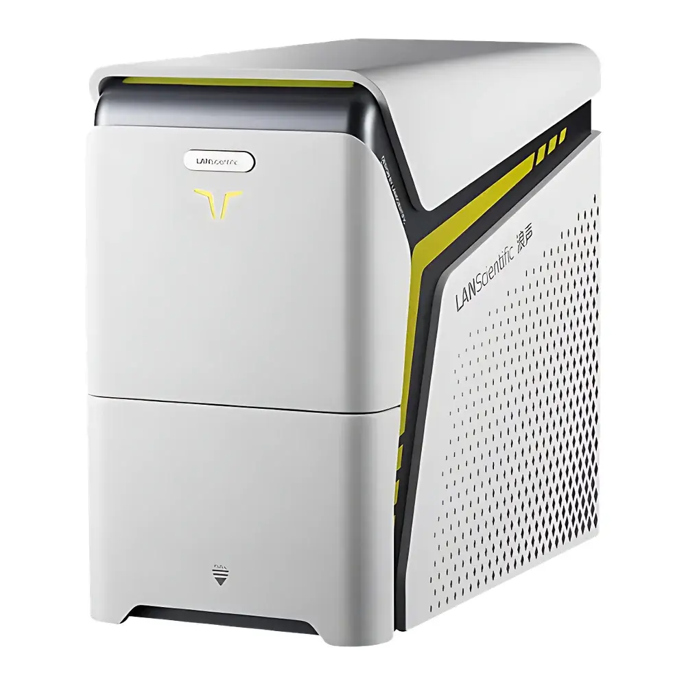
Brookfield LANScientific SuperSEM N10 Desktop Scanning Electron Microscope
| Brand | LANScientific |
|---|---|
| Origin | Jiangsu, China |
| Manufacturer Type | Direct Manufacturer |
| Origin Category | Domestic (China) |
| Model | SuperSEM N10 |
| Instrument Type | Benchtop SEM |
| Electron Source | Tungsten Filament |
| SEM Class | Entry-Level Tungsten-Filament SEM |
| Max Sample Diameter | 90 mm |
| Max Sample Thickness | 40 mm |
| Stage Travel | X: 25 mm, Y: 25 mm, Z: 30 mm |
Overview
The Brookfield LANScientific SuperSEM N10 is a compact, benchtop scanning electron microscope engineered for precision surface morphology imaging and integrated energy-dispersive X-ray spectroscopy (EDS) elemental analysis. Utilizing a thermionic tungsten filament electron source, the system delivers stable beam current and consistent landing energy—optimized for routine characterization of conductive and semi-conductive samples without requiring ultra-high vacuum or cryogenic cooling. Its modular electron optical column incorporates a dual-detector architecture (secondary electron and high-sensitivity quad-segmented backscattered electron detectors), enabling simultaneous topographic and compositional contrast generation. Unlike conventional standalone EDS add-ons, the SuperSEM N10 embeds real-time spectral acquisition and processing directly into the imaging workflow—leveraging synchronized BSE video streaming and EDS pulse processing to generate quantitative elemental maps during active scanning. Designed for laboratories with space constraints and moderate throughput requirements, it operates under standard 220 V/50 Hz mains power and requires no dedicated vibration isolation platform or HVAC-controlled environment.
Key Features
- Integrated real-time EDS capability: Simultaneous acquisition of BSE video and characteristic X-ray spectra enables live elemental mapping without interrupting imaging.
- Quad-segmented backscattered electron (BSE) detector: Provides angular-selective signal separation for enhanced atomic number contrast and phase discrimination.
- Automated operation suite: One-click auto-focus, auto-stigmation, auto-contrast/brightness, and large-area image stitching reduce operator dependency and training time.
- High-bandwidth signal acquisition: 10 MHz digitization bandwidth supports fast scanning speeds with minimal motion artifacts—ideal for dynamic observation in video mode.
- Infrared CCD navigation camera: Enables rapid sample positioning and real-time chamber monitoring without breaking vacuum.
- Compact footprint (< 0.8 m²): Fits on standard lab benches; compatible with standard electrical outlets and ambient laboratory conditions.
Sample Compatibility & Compliance
The SuperSEM N10 accommodates specimens up to 90 mm in diameter and 40 mm in thickness, with a motorized three-axis stage offering 25 mm (X), 25 mm (Y), and 30 mm (Z) travel. Conductive samples require no coating; non-conductive materials may be analyzed with optional low-vacuum mode or light carbon sputter coating. The system complies with IEC 61000-6-3 (EMC emissions) and IEC 61000-6-2 (immunity) standards. While not certified for GMP/GLP environments out-of-the-box, its software audit trail functionality—including timestamped parameter logs, user session records, and raw data export—supports alignment with ISO/IEC 17025 documentation requirements for testing laboratories.
Software & Data Management
The proprietary SuperSEM Control Suite features an intuitive graphical interface built on Qt framework, supporting Windows 10/11 64-bit OS. It includes real-time spectral overlay, live stoichiometric quantification (ZAF correction), and pseudo-color EDS mapping with adjustable ROI thresholds. All acquired images, spectra, and metadata are stored in vendor-neutral formats (TIFF, CSV, .spc). Raw detector signals and stage coordinates are logged at acquisition time, enabling retrospective reprocessing. Software version history and configuration snapshots are retained for traceability—facilitating internal validation protocols and regulatory review readiness per FDA 21 CFR Part 11 when deployed with appropriate IT governance controls.
Applications
- Materials Science: Phase identification in alloys, ceramic grain boundary analysis, coating uniformity assessment, and battery electrode microstructure evaluation.
- Geoscience: Mineral assemblage mapping in thin sections, porosity quantification in sedimentary rocks, and volcanic ash particle morphology studies.
- Life Sciences: Morphological examination of dried biological tissues, pollen grain surface texture analysis, and biomaterial scaffold characterization.
- Forensics: Gunshot residue (GSR) particle detection, fiber comparison, and trace evidence elemental fingerprinting.
- Quality Control: Defect root-cause analysis in electronics packaging, weld seam inspection, and additive manufacturing powder morphology screening.
FAQ
What vacuum level does the SuperSEM N10 operate at?
The system maintains a typical operating pressure of 1 × 10⁻³ Pa in high-vacuum mode, achieved via a turbomolecular pump backed by a dry scroll pump.
Is conductive coating required for non-metallic samples?
For optimal secondary electron imaging, non-conductive samples should be coated with ~5–10 nm carbon or gold using a sputter coater; optional low-vacuum mode permits limited uncoated imaging of hydrated or insulating specimens.
Can EDS spectra be acquired simultaneously with SE/BSE imaging?
Yes—the instrument synchronizes detector readout across all channels, allowing concurrent collection of topographic, compositional, and spectral data during a single scan pass.
Does the software support batch processing of multiple EDS maps?
The Control Suite includes scripting support (Python API) for automated map alignment, normalization, and statistical reporting across multi-sample datasets.
What maintenance intervals are recommended for the tungsten filament?
Filament lifetime averages 150–200 hours under typical operating conditions (20 kV, 1 nA); replacement is a field-serviceable procedure requiring < 30 minutes and no realignment tools.


