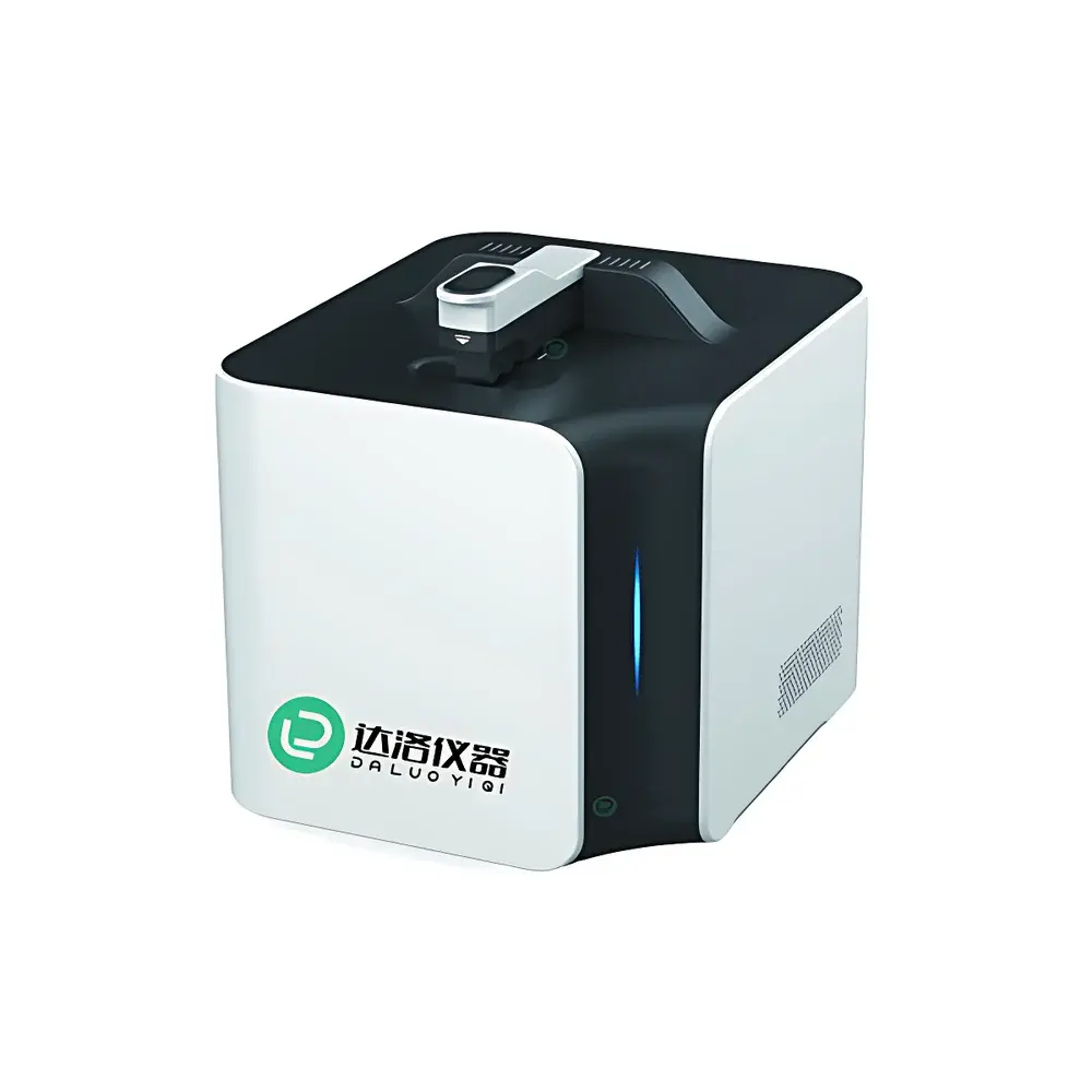
Daxluot-MC Automated Cell Counter
| Brand | Daxluot |
|---|---|
| Origin | Shanghai, China |
| Model | Daxluot-MC |
| Detection Time (Brightfield) | <3 sec |
| Cell Concentration Range | 7×10²–2.5×10⁷ cells/mL |
| Adjustable Sample Height | 50 µm (5 µL), 100 µm (10 µL), 400 µm (40 µL) |
| Cell Diameter Range | 4–400 µm |
| Imaging Resolution | 5 MP brightfield |
| Excitation/Filter | AO 530 nm, PI 620 nm |
| Illumination | LED-based brightfield |
| Sample Stage Material | Hydrophilic quartz glass |
| Focus | Auto- and manual-focus modes |
| Power Supply | 12 V DC |
Overview
The Daxluot-MC Automated Cell Counter is an optical image-based cell analysis instrument engineered for precision, reproducibility, and operational simplicity in routine cell culture workflows. It employs high-resolution brightfield microscopy combined with fluorescence imaging capabilities (acridine orange / propidium iodide and GFP channels) to deliver quantitative metrics—including total cell concentration, viable cell count, viability percentage, and average cell diameter—without requiring disposable counting chambers. Unlike traditional hemocytometer-based or fixed-height automated counters, the Daxluot-MC implements variable-depth sample chamber technology: users select one of three calibrated chamber heights (50 µm, 100 µm, or 400 µm) to match sample concentration and cell size, thereby optimizing signal-to-noise ratio and minimizing counting bias across heterogeneous or low-abundance suspensions. The system utilizes a hydrophilic quartz glass sample stage that ensures uniform capillary loading and minimal surface tension artifacts, while its 5-megapixel sensor captures high-fidelity images suitable for post-acquisition review and audit compliance.
Key Features
- Variable-depth sample chamber: Three user-selectable chamber heights (50 µm, 100 µm, 400 µm) enable accurate enumeration across broad concentration ranges—from sparse primary isolates to dense suspension cultures.
- Multi-modal detection: Supports brightfield, trypan blue exclusion, AO/PI dual-fluorescence, and GFP-compatible imaging—all on a single platform without hardware reconfiguration.
- Rapid analysis: Brightfield cell counting completed in under 3 seconds per sample; fluorescence mode processing time remains consistent with standard image acquisition and segmentation algorithms.
- Chamber-free operation: Eliminates dependency on consumable counting slides or microfluidic cartridges, reducing per-test cost and waste generation.
- Intelligent focusing: Dual-mode autofocus algorithm optimized for cell monolayers and clustered aggregates, supplemented by manual fine-tuning capability for edge-case samples (e.g., highly viscous media or debris-rich preparations).
- Optical architecture: LED-based brightfield illumination at 530 nm with matched emission filters (AO: 530 nm bandpass; PI: 620 nm longpass) ensures spectral fidelity and minimal phototoxicity during repeated imaging cycles.
Sample Compatibility & Compliance
The Daxluot-MC accommodates a wide spectrum of mammalian, insect, yeast, and plant-derived cells—including adherent lines passaged via enzymatic or mechanical dissociation—as well as primary isolates such as PBMCs, hepatocytes, and neural stem cells. Its 4–400 µm detection range supports both small lymphocytes and large megakaryocytes or spheroids. Sample volumes are configurable between 5 µL and 40 µL, making it compatible with low-yield biopsies and micro-scale culture formats (e.g., 96-well plate harvests). While not certified to ISO 13485 or FDA 21 CFR Part 11 out-of-the-box, the instrument’s data export structure (CSV, TIFF, JPEG) and timestamped image metadata support integration into GLP/GMP-aligned laboratory information management systems (LIMS) when paired with validated SOPs. Raw image archives satisfy internal audit requirements for traceability in QC/QA environments.
Software & Data Management
The embedded software provides real-time image preview, adjustable segmentation thresholds, and batch-processing capability for multi-sample runs. All acquired images are stored with embedded EXIF metadata—including acquisition time, chamber height, exposure settings, and user ID—and exported in lossless TIFF format for third-party analysis (e.g., ImageJ, MATLAB, or Python-based morphometrics). Quantitative outputs include cell concentration (cells/mL), viability (%), mean diameter (µm), coefficient of variation (CV%), and population histograms. Exported CSV files conform to ANSI X12 and ASTM E1461-22 data formatting conventions for interoperability with ELN platforms. No cloud connectivity or remote telemetry is enabled by default; all data resides locally unless explicitly transferred via encrypted USB or network share.
Applications
- Bioprocess monitoring: Daily viability and density tracking during upstream bioreactor runs or shake flask expansions.
- Vaccine and viral vector production: Quantification of HEK293 or Sf9 host cells pre-transfection and post-harvest.
- Cell therapy QC: Release testing of CAR-T or MSC products per USP sterility and viability criteria.
- Academic research: Longitudinal studies of cytotoxicity, proliferation kinetics, or CRISPR editing efficiency using AO/PI or GFP reporters.
- Stem cell labs: Assessment of embryoid body dissociation efficiency and single-cell recovery yield.
FAQ
Does the Daxluot-MC require calibration with standardized beads or reference standards?
No—system calibration is factory-performed using NIST-traceable micrometer standards; users perform only routine verification using supplied control suspensions.
Can the instrument distinguish between cell doublets and true single cells?
Yes—the 5 MP resolution and adaptive thresholding algorithm resolve intercellular boundaries down to ~2 µm separation, enabling reliable doublet discrimination for cells >8 µm in diameter.
Is the quartz sample stage autoclavable or chemically resistant?
The hydrophilic quartz surface withstands 70% ethanol, isopropanol, and 0.1% SDS cleaning; however, autoclaving is not recommended due to thermal stress risk on bonded optical components.
What file formats are supported for image export?
TIFF (uncompressed, 16-bit), JPEG (high-quality), and CSV (tabular results with metadata headers).
How is data integrity ensured during power interruption?
All image acquisition and analysis steps are transactionally logged; incomplete runs are flagged and recoverable upon reboot without data loss.


