Evident SLIDEVIEW VS200 SILA Ultra-Fast Optical Sectioning Whole-Slide Imaging System
| Brand | Evident (formerly Olympus) |
|---|---|
| Origin | Japan |
| Manufacturer Type | Original Equipment Manufacturer (OEM) |
| Import Status | Imported Instrument |
| Model | VS200 SILA |
| Medical Device Classification | Non-Medical Device |
| Instrument Class | Research-Grade Fluorescence Microscope |
Overview
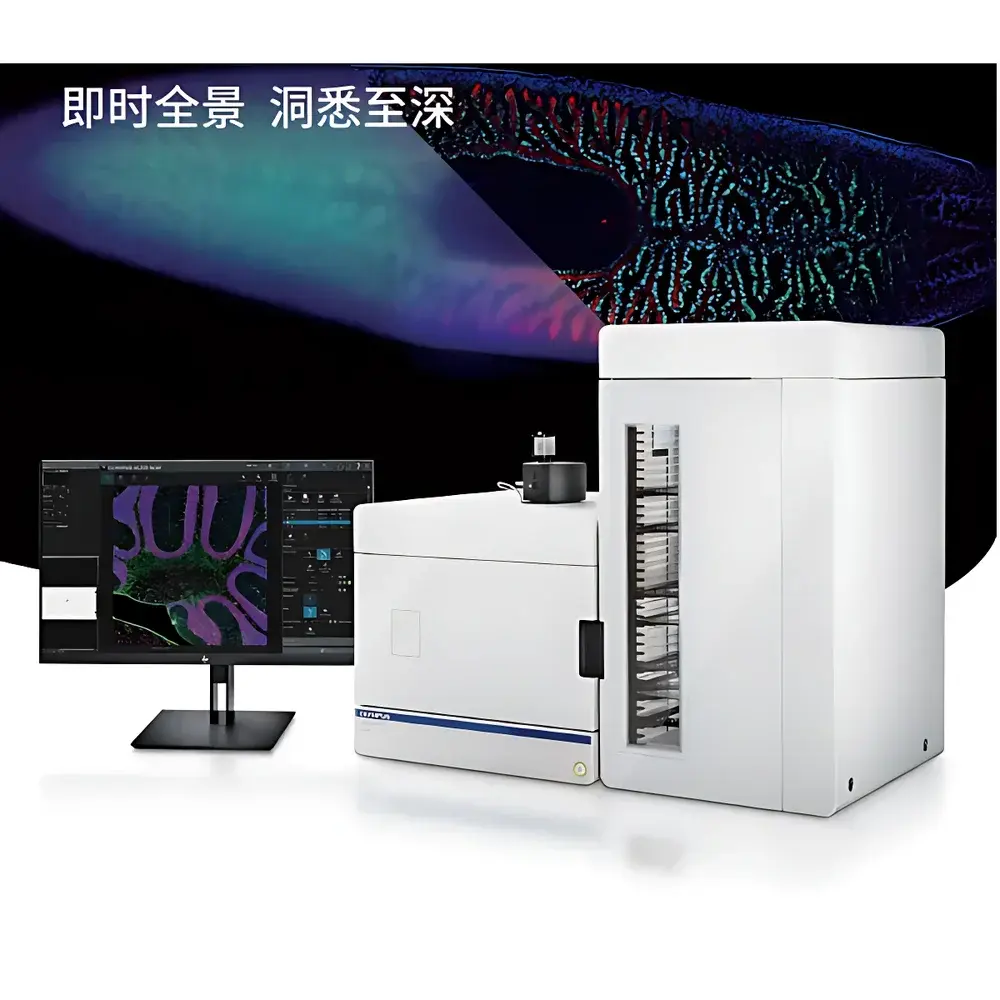
The Evident SLIDEVIEW VS200 SILA is a high-performance, research-grade whole-slide imaging (WSI) system engineered for ultra-fast optical sectioning of thick biological specimens without mechanical Z-stacking. Unlike conventional widefield scanners—whose out-of-focus blur degrades contrast and resolution in samples >20 µm—or point-scanning confocal systems—whose serial acquisition limits throughput—the VS200 SILA employs structured illumination with laser-based speckle pattern projection and real-time computational extraction of in-focus signal layers. This approach implements optical sectioning principles analogous to confocal microscopy but at camera-based parallel acquisition speeds, enabling true 3D volumetric reconstruction from single-pass scanning. The system is optimized for neuroscience (e.g., 30–100 µm brain sections), organoid imaging, plant tissue mounts, and developmental biology specimens where axial clarity, large-field fidelity, and quantitative reproducibility are critical. Its architecture supports both fixed and live-cell compatible protocols (non-invasive illumination intensity control), and it operates under standard laboratory environmental conditions without requiring vibration isolation or climate-controlled enclosures.
Key Features
- Optical sectioning capability equivalent to confocal resolution (effective axial resolution ~1.5–2.0 µm) without Z-stack acquisition or post-processing deconvolution
- Simultaneous multi-modal imaging: brightfield, fluorescence (widefield and optical-sectioned), darkfield, phase contrast, and simple polarized light
- High-throughput automated loader accommodating up to 210 standard (26 × 76 mm) or oversized slides (up to 102 × 127 mm) per batch with mixed-size and mixed-stain compatibility
- Single-parameter optimization: users adjust only the optical sectioning thickness (1–10 µm range) to balance depth discrimination and signal-to-noise ratio
- Real-time focal plane extraction using proprietary hardware-accelerated algorithms; no external GPU or post-scan computing required
- Integrated TruAI deep learning framework supporting user-trained CNN models for ROI detection (e.g., pancreatic islets, glomeruli, tumor foci) and selective high-resolution scanning
Sample Compatibility & Compliance
The VS200 SILA accommodates diverse specimen formats including paraffin-embedded and frozen tissue sections (10–100 µm), cleared whole-mounts (CLARITY, iDISCO), organoids, spheroids, and plant histological preparations. It complies with ISO 13485 design control principles for research instrumentation and meets electromagnetic compatibility (EMC) requirements per IEC 61326-1. While not classified as a medical device under FDA 21 CFR Part 809 or EU IVDR, its output data structure conforms to DICOM Supplement 145 (Whole Slide Imaging) and OpenSlide-compatible TIFF pyramids, ensuring interoperability with PACS and digital pathology platforms used in translational research environments. All firmware and software modules support audit trail logging per GLP/GMP-aligned documentation practices, including timestamped operator actions, parameter changes, and scan metadata export.
Software & Data Management
SLIDEVIEW Software v4.0 provides unified control, acquisition, and visualization workflows. It natively generates multi-resolution pyramid TIFF files compliant with OME-TIFF specifications and supports direct export to cloud-based analysis platforms (e.g., QuPath, HALO, Visiopharm). The TruAI module enables model import/export in ONNX format and includes built-in tools for annotation, training dataset curation, and inference validation. Scan metadata—including objective magnification, excitation/emission filter sets, exposure time, sectioning thickness, and stage position—is embedded in EXIF and OMEXML headers. Data integrity is ensured via SHA-256 checksum generation during export and optional integration with LIMS via RESTful API endpoints. No proprietary file locking or vendor-specific viewer dependency is imposed.
Applications
The VS200 SILA serves core use cases across preclinical and basic research domains:
- Neuroanatomy mapping: high-fidelity 3D reconstruction of immunolabeled mouse brain sections (e.g., DAPI/GFAP/Iba1/NeuN multiplex) for regional cell density quantification
- Tumor microenvironment analysis: spatial profiling of immune infiltrates, stromal interfaces, and vascular networks in thick FFPE tumor sections
- Developmental biology: time-resolved imaging of embryonic organogenesis in optically cleared specimens
- Plant science: layer-resolved imaging of vascular bundles, stomatal complexes, and fungal colonization in leaf cross-sections
- Educational digitization: creation of interactive, zoomable slide libraries for histology teaching with embedded annotations and layered channel overlays
FAQ
Does the VS200 SILA require specialized training to operate?
No formal certification is required. Basic operation involves loading slides, selecting objective and mode, setting sectioning thickness, and initiating scan—typically mastered within one hour by lab personnel familiar with standard microscope controls.
Can the system perform true confocal-level Z-stack acquisition?
No—it does not acquire sequential optical sections mechanically. Instead, it delivers equivalent axial resolution and contrast through single-pass optical sectioning, eliminating the need for time-consuming Z-series collection.
Is AI model training performed on-device or externally?
Model training occurs externally using Evident’s TruAI Studio (Windows/Linux); trained models (.onnx) are imported into the VS200 SILA for inference-only deployment during scanning.
What is the maximum supported slide thickness for optical sectioning?
Specimens up to 100 µm thick have been validated in peer-reviewed studies; optimal performance is observed between 20–60 µm depending on refractive index homogeneity and labeling density.
Does the system support live-cell imaging?
It supports short-term (<30 min), low-light live imaging of fluorescently labeled cells or tissues under ambient CO₂ and temperature control when integrated with environmental chambers—but is not designed for long-term time-lapse microscopy.
Related Products


