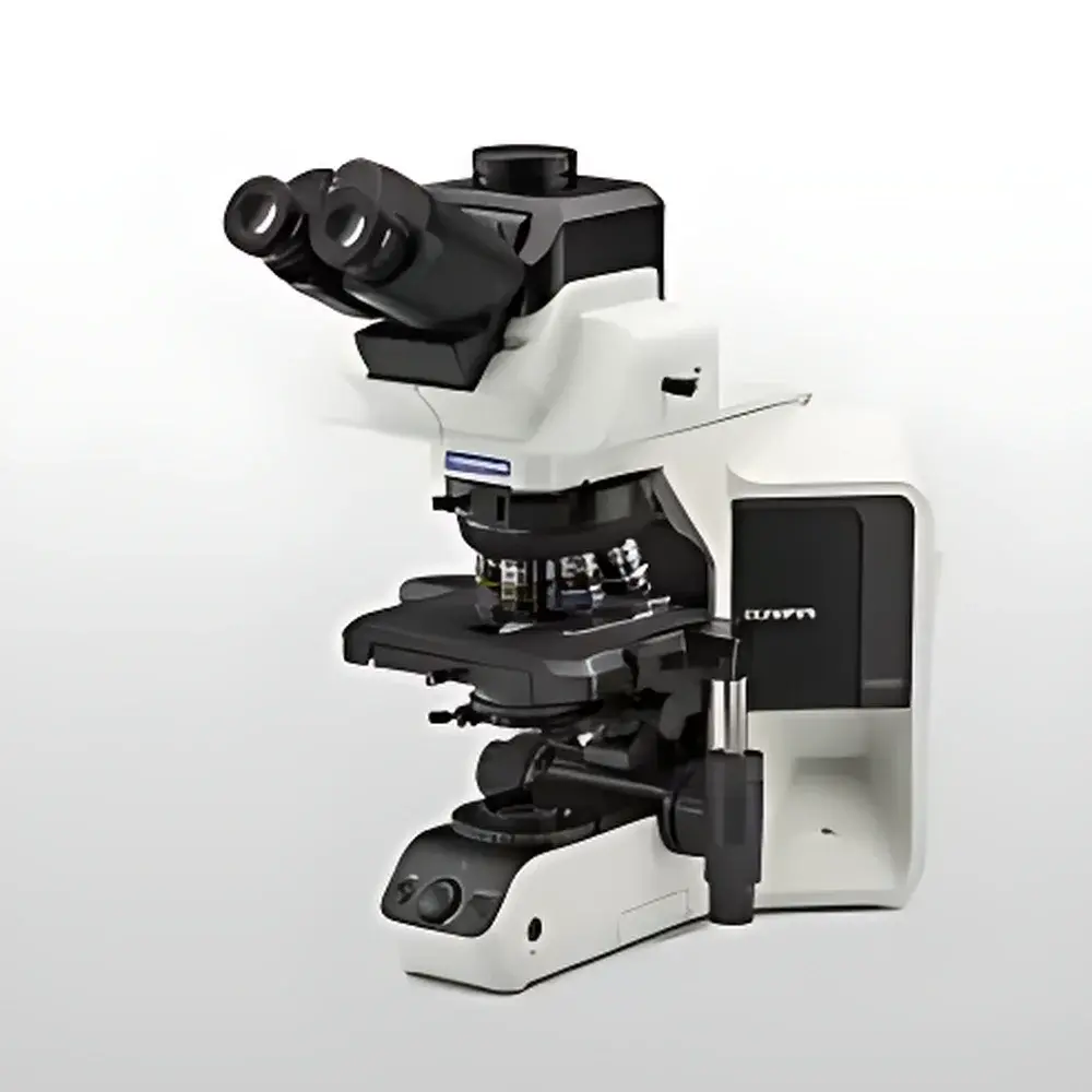
OLYMPUS BX53(LED) Semi-Motorized Fluorescence Microscope
| Brand | OLYMPUS |
|---|---|
| Origin | Japan |
| Manufacturer Type | Original Equipment Manufacturer (OEM) |
| Origin Category | Imported |
| Model | BX53(LED) |
| Instrument Type | Upright Fluorescence Microscope |
| Excitation Source | High-Intensity LED |
| Instrument Class | Conventional Fluorescence Microscope |
| Objective Options | Multiple X Line Objectives (e.g., 4×, 10×, 20×, 40×, 60×, 100× oil) |
| Fluorescence Modules | Interchangeable Filter Cube Turrets (e.g., FITC, TRITC, DAPI, Cy5) |
| Illumination | 100 W Mercury Arc Lamp (optional), Fiber-Optic Light Guide (optional), Integrated LED Illuminator (standard) |
| Control Mode | Manual and Motorized |
| Focusing Mechanism | Coaxial Fine/Coarse Focus with Motorized Z-Drive Option |
| Condenser Options | U-UCD8, U-LC, U-ULC, U-SC3 |
| Observation Heads | Binocular or Trinocular (with C-mount port) |
| DIC Components | Available as Add-on Module (U-DICR, U-DICB) |
| Stage | Mechanical XY Stage (76 × 52 mm travel), Optional Motorized Stage |
| Fluorescence Filter Sets | Broadband and Narrowband Bandpass Filters, Dichroic Mirrors, Emission Filters (all standardized to ISO 10934-1) |
Overview
The OLYMPUS BX53(LED) Semi-Motorized Fluorescence Microscope is an upright, modular optical platform engineered for high-fidelity fluorescence imaging in academic research, clinical pathology, and cell biology laboratories. Built upon Olympus’ proven BX optical architecture, the system integrates a high-stability, vibration-damped mechanical frame with precision-machined optical pathways optimized for Köhler illumination and minimal chromatic aberration. Its core fluorescence measurement principle relies on epifluorescence excitation via LED-based solid-state illumination—delivering stable, flicker-free output with spectral fidelity closely matching traditional mercury arc sources (particularly in the 365–570 nm range), while eliminating UV ozone generation and thermal drift associated with arc lamps. The microscope supports both widefield fluorescence and optional Differential Interference Contrast (DIC), enabling label-free structural visualization alongside fluorescent labeling. Designed for reproducibility in regulated environments, the BX53(LED) complies with ISO 9001 manufacturing standards and meets essential safety requirements per IEC 61010-1 for laboratory electrical equipment.
Key Features
- Integrated high-output LED illuminator with adjustable intensity control (0–100%), offering >10,000-hour lifetime and negligible heat emission at the specimen plane—critical for live-cell imaging and long-term time-lapse experiments.
- X Line objective series featuring apochromatic correction across visible and near-UV spectra, flat-field imaging over ≥90% of the field of view, and enhanced numerical aperture (NA up to 1.45) for superior resolution and signal-to-noise ratio in fluorescence detection.
- Semi-motorized configuration: motorized nosepiece (with optional encoder for automatic magnification registration), motorized filter cube turret (6-position), and motorized condenser height adjustment—enabling repeatable setup recall and protocol-driven acquisition.
- Modular optical path design supporting simultaneous or sequential fluorescence, phase contrast, DIC, and brightfield observation without realignment—facilitating multi-modal correlative imaging workflows.
- Trinocular head with dedicated C-mount port (1× or 0.5× reduction) ensures optimal coupling with scientific CMOS or sCMOS cameras; all optical components are anti-reflection coated to minimize stray light and maximize transmission efficiency above 92%.
- Ergonomic stage and focus controls conform to ISO 14738 industrial ergonomics guidelines, reducing operator fatigue during extended microscopy sessions.
Sample Compatibility & Compliance
The BX53(LED) accommodates standard glass slides (26 × 76 mm), petri dishes (up to 100 mm diameter), multi-well plates (6–96-well), and custom chambered coverslips. Its high-precision mechanical stage enables accurate coordinate mapping for serial section alignment and tissue microarray analysis. All fluorescence filter cubes meet ISO 10934-1 spectral tolerances for center wavelength (±2 nm), bandwidth (±5%), and blocking depth (>OD6 beyond passband). For regulatory compliance, the system supports audit-trail-enabled imaging protocols when paired with cellSens software under FDA 21 CFR Part 11-compliant configurations (electronic signatures, user access control, versioned metadata logging). It is routinely deployed in GLP-compliant histopathology labs and ISO/IEC 17025-accredited testing facilities.
Software & Data Management
The microscope is fully compatible with Olympus cellSens Dimension software (v3.1+), a validated platform for quantitative fluorescence analysis, Z-stack reconstruction, deconvolution, and multi-channel co-localization. cellSens supports DIC vector analysis, intensity profiling along user-defined lines, and batch processing with scripting (Python API). Raw image data is saved in TIFF or OIR (Olympus Image Repository) format with embedded EXIF metadata—including objective ID, filter set, exposure time, LED intensity, and stage coordinates. Integration with third-party platforms (e.g., ImageJ/Fiji via Bio-Formats, MATLAB via SDK) is supported through open file I/O and COM automation interfaces. All software updates and calibration utilities are distributed via Olympus’ secure firmware portal with SHA-256 checksum verification.
Applications
- Clinical cytology and histopathology: rapid screening of Papanicolaou smears, immunofluorescent staining of FFPE tissue sections, and dual-label detection of biomarkers (e.g., ER/PR in breast cancer diagnostics).
- Live-cell dynamics: time-lapse tracking of GFP-tagged cytoskeletal proteins under low-phototoxicity LED illumination, combined with DIC for membrane topology reference.
- Neuroscience: high-resolution imaging of dendritic spines in organotypic brain slices using Alexa Fluor 488/594 conjugated antibodies and structured illumination-compatible optics.
- Microbiology: detection of autofluorescent bacterial aggregates (e.g., Pseudomonas aeruginosa pyocyanin) and viability assessment via SYTO9/PI dual staining.
- Materials science: fluorescence-assisted characterization of quantum dot-labeled polymer composites and photoluminescent nanomaterials.
FAQ
Is the BX53(LED) compatible with third-party fluorescence cameras?
Yes—the trinocular port supports industry-standard C-mount and F-mount adapters; sensor size compatibility extends from 1/2″ to 4/3″ formats, with full electronic trigger and TTL synchronization support.
Can the LED illuminator be calibrated for quantitative intensity measurements?
Yes—Olympus provides NIST-traceable irradiance calibration kits (Cat. No. U-LEDCAL) for absolute photon flux quantification at the specimen plane, valid across all installed filter sets.
Does the system support automated Z-stack acquisition with hardware autofocus?
While the base BX53(LED) lacks integrated hardware autofocus, it supports external piezo Z-drives (e.g., Physik Instrumente P-725) via analog voltage input and cellSens hardware synchronization modules.
Are DIC prisms and Nomarski components included by default?
No—DIC modules (U-DICR, U-DICB) and matching Wollaston prisms are optional accessories; they require corresponding condenser (U-ULC) and objective-specific shear adjustments.
What maintenance intervals are recommended for the LED illumination system?
No scheduled lamp replacement is required; however, annual photometric verification using a calibrated spectroradiometer is recommended to ensure spectral stability and intensity linearity per ISO/IEC 17025 internal quality procedures.


