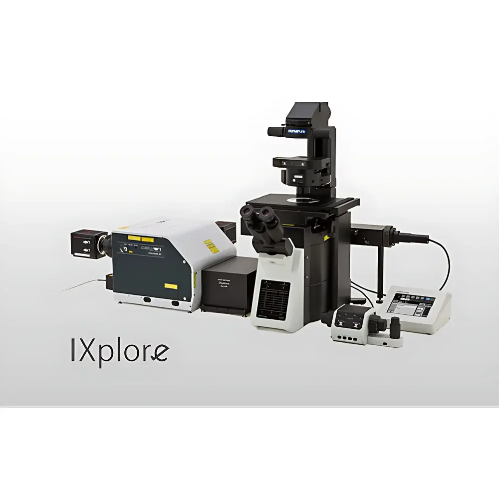
Olympus IXplore SpinSR Spinning-Disk Confocal Super-Resolution Microscope
| Brand | Olympus |
|---|---|
| Origin | Japan |
| Manufacturer Type | Original Equipment Manufacturer (OEM) |
| Origin Category | Imported |
| Model | IXplore SpinSR |
| Pricing | Available Upon Request |
Overview
The Olympus IXplore SpinSR is a high-performance spinning-disk confocal super-resolution microscope engineered for quantitative, live-cell imaging with minimal phototoxicity and photobleaching. It integrates conventional widefield fluorescence, standard confocal, and optical super-resolution modalities—specifically Olympus Super-Resolution (OSR)—into a single, unified platform based on structured illumination and multi-frame deconvolution principles. Unlike point-scanning confocal systems, the SpinSR employs a dual-disk architecture (excitation and emission) to enable parallelized photon collection across the field of view, delivering high-speed volumetric imaging while preserving cellular physiology over extended time-lapse experiments. Its optical design supports true 3D reconstruction with sub-diffraction spatial resolution down to 120 nm in the XY plane and ~350 nm axially—achieved without requiring specialized fluorophores or single-molecule localization techniques. The system is built upon the IXplore inverted microscope platform, ensuring mechanical stability, precise motorized Z-control (with 0.01 µm step resolution), and compatibility with environmental chambers for long-term physiological imaging.
Key Features
- Spinning-disk confocal architecture enabling high-speed, low-phototoxicity imaging at up to 100 frames per second (full frame, 2048 × 2048 pixels) under optimal conditions
- Olympus Super-Resolution (OSR) mode delivering consistent 120 nm XY resolution via computational enhancement of confocal z-stacks, validated against NIST-traceable fluorescent bead standards
- Seamless, software-controlled switching among widefield, confocal, and OSR imaging modes—no hardware reconfiguration required
- Silicone oil immersion objectives (e.g., UPLSAPO 60×/1.30 SI) minimizing spherical aberration across thick specimens and enabling accurate 3D reconstruction with axial fidelity
- Integrated TruSight multicolor processing engine supporting spectral unmixing and crosstalk correction for ≥4-channel acquisition without dedicated STED or PALM dyes
- Real-time GPU-accelerated deconvolution using Olympus’ proprietary algorithms, improving signal-to-noise ratio and contrast without compromising temporal resolution
Sample Compatibility & Compliance
The IXplore SpinSR supports a broad range of biological specimens—from adherent monolayers (e.g., HeLa, primary neurons) to organoids, spheroids, and live zebrafish embryos—under controlled CO₂, temperature, and humidity environments. Its low-light sensitivity (quantum efficiency >85% at 520 nm) and high dynamic range (>28,000:1) ensure robust detection of endogenous or weakly expressed fluorescent reporters (e.g., GFP-EB3, mCherry-tagged histones). The system complies with ISO 10993-5 (biological evaluation of medical devices) for optical components in contact with cell culture media, and its firmware architecture supports audit trails and electronic signatures in accordance with FDA 21 CFR Part 11 for regulated research environments. All imaging protocols are compatible with GLP/GMP-aligned documentation workflows when integrated with Olympus cellSens software.
Software & Data Management
Controlled exclusively through Olympus cellSens Dimension v3.x or later, the SpinSR provides deterministic acquisition scripting, batch processing pipelines, and metadata-rich TIFF/HDF5 export. The software includes built-in tools for colocalization analysis (Pearson’s r, Manders’ coefficients), 3D surface rendering, spine morphometry (for neuronal applications), and time-series registration using intensity-based rigid-body alignment. Raw OSR datasets are stored with embedded calibration parameters (e.g., pixel size, Z-step, objective NA), enabling reproducible reprocessing. Data integrity is preserved via checksum validation during transfer and optional integration with LIMS or ELN systems via RESTful API.
Applications
- Long-term live-cell mitosis tracking—visualizing kinetochore–microtubule dynamics with <2-min temporal resolution and sub-150 nm structural discrimination
- 3D time-lapse imaging of dendritic spine maturation in co-cultured mouse primary neurons and astrocytes, resolving immature vs. mature spine morphology over 1-hour acquisitions (41 Z-slices @ 0.15 µm steps)
- High-content screening of ciliary ultrastructure (e.g., Odf2 localization in basal bodies) in epithelial models under physiological shear stress
- Quantitative nuclear pore complex (NPC) mapping—simultaneous dual-color OSR imaging of Nup153 and Nup62 with <5 nm lateral registration accuracy
- Fixed-tissue super-resolution cytology—multi-target immunofluorescence of spindle apparatus components (α-tubulin, Hec1, DAPI) without antibody cross-reactivity artifacts
FAQ
What is the effective resolution limit of the IXplore SpinSR in OSR mode?
The system achieves a verified lateral resolution of ≤120 nm (FWHM) in XY and ≤350 nm in Z, as measured using sub-resolution fluorescent beads and confirmed by Fourier ring correlation (FRC) analysis.
Does OSR require special fluorophores or buffer conditions?
No—OSR operates with conventional organic dyes (e.g., Alexa Fluor 488/555/647) and standard aqueous mounting media; no oxygen scavenging or photoswitching buffers are needed.
Can the SpinSR be integrated into automated core facilities?
Yes—it supports remote operation via TCP/IP, scheduled acquisition, and third-party scheduler compatibility (e.g., MetaXpress, Acapella) through standardized device control protocols.
Is the silicone oil immersion objective compatible with live-cell imaging?
Yes—the UPLSAPO 60×/1.30 SI objective maintains stable refractive index matching during prolonged imaging (>4 hours) and is certified for use with commercial environmental chambers.
How does the SpinSR handle chromatic aberration across multicolor experiments?
The system incorporates hardware-based spectral calibration (using reference lasers) and software-driven channel alignment, achieving <0.05 µm residual offset across 405–780 nm excitation bands.


