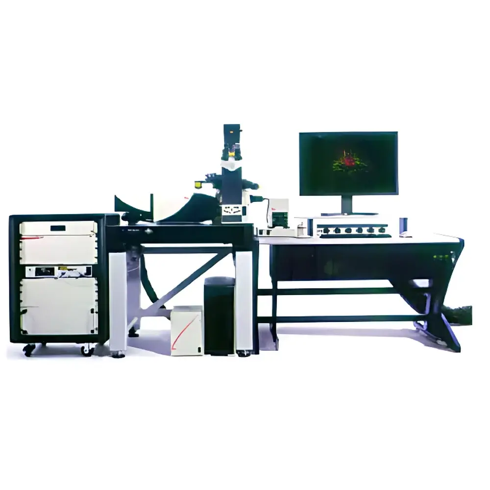
Leica TCS SP8 Laser Scanning Confocal Microscope
| Brand | Leica |
|---|---|
| Origin | Germany |
| Model | TCS SP8 |
| Type | Laser Scanning Confocal Microscope |
| Optical Architecture | Upright or Inverted Configurable Platform |
| Detection Principle | Point-scanning confocal fluorescence imaging with spectral detection and time-gated photon counting capability |
| Maximum Frame Rate | 428 fps (at 512 × 32 resolution) |
| Field of View | Up to 22 mm diagonal (with resonant scanner and wide-field optics) |
| Scan Modes | Galvoscanner, Resonant Scanner, Galvoflow (combined galvo-resonant hybrid mode for accelerated Z-stack acquisition) |
| Detector Options | Hybrid Detectors (HyD), PMTs, Spectral Detectors (Tuneable Bandpass or Linear Unmixing) |
| Excitation Sources | Integrated white-light laser (WLL, 470–670 nm), optional UV/IR lasers, acousto-optic tunable filters (AOTF) |
| Software Platform | LAS X (v3.7+), compliant with FDA 21 CFR Part 11 audit trail and electronic signature modules |
| Compliance | CE, IEC 61000-6-3, ISO 13485 (for medical device configurations), GLP/GMP-ready metadata logging |
Overview
The Leica TCS SP8 Laser Scanning Confocal Microscope is an advanced point-scanning fluorescence imaging platform engineered for quantitative, high-fidelity optical sectioning in live and fixed biological specimens. It operates on the fundamental principle of spatially filtered detection: a focused laser beam scans across the specimen pixel-by-pixel, while a conjugate pinhole rejects out-of-focus emission photons—enabling true optical sectioning with sub-micron axial resolution (~0.5 µm at 550 nm emission). Its optical architecture integrates a tunable white-light laser (470–670 nm), acousto-optic tunable filters (AOTF), and hybrid detectors (HyD) optimized for single-photon sensitivity and low-noise signal acquisition. The system’s photon efficiency—achieved through proprietary light path design, anti-reflective coated optics, and minimized internal scattering—is validated by >30% higher signal-to-noise ratio (SNR) compared to prior-generation confocals under identical illumination conditions. This enables prolonged live-cell imaging with reduced phototoxicity and photobleaching, supporting long-term timelapse studies of dynamic subcellular processes without compromising viability.
Key Features
- Galvoflow scanning mode: Synchronizes galvanometric and resonant scanners to accelerate Z-stack acquisition by up to 3× while maintaining confocal resolution and minimizing stage-induced drift.
- 22 mm field-of-view capability: Achieved via optimized scan optics and wide-field tube lens configuration—enabling high-resolution mosaic imaging of large tissue sections or multi-well plates without mechanical tiling artifacts.
- Spectral detection with linear unmixing: Captures full emission spectra (350–900 nm) at each pixel, allowing robust separation of spectrally overlapping fluorophores (e.g., GFP/mCherry/Cy5) without filter wheel exchange.
- Time-gated detection (optional): Supports fluorescence lifetime imaging (FLIM) integration for molecular environment sensing—compatible with pulsed lasers and TCSPC modules.
- Modular optical path: Supports upright and inverted configurations; interchangeable objectives (including water-dipping, silicone-oil, and long-working-distance lenses); motorized filter turrets and focus drives with nanometer-scale repeatability.
- Environmental chamber compatibility: Designed for integration with CO₂, temperature, and humidity control systems—validated for sustained imaging at 37°C ± 0.2°C over 72-hour periods.
Sample Compatibility & Compliance
The TCS SP8 accommodates diverse sample formats including glass-bottom dishes (thickness #1.5), multi-well plates (6–384-well), organotypic slices, cleared tissues (CLARITY, iDISCO), and whole-mount embryos. Its low-phototoxicity operation supports sensitive models such as primary neurons, zebrafish embryos, and human induced pluripotent stem cell (iPSC)-derived organoids. Regulatory compliance includes CE marking per Directive 2014/30/EU (EMC) and 2014/35/EU (LVD); ISO 13485 certification applies to configurations used in IVD research and preclinical assay development. All acquired datasets embed FAIR-compliant metadata (OME-TIFF format), and LAS X software supports 21 CFR Part 11–compliant user access controls, electronic signatures, and immutable audit trails—meeting GLP and GMP documentation requirements for regulated laboratories.
Software & Data Management
LAS X software (v3.7 or later) serves as the unified acquisition, analysis, and reporting engine. It provides real-time spectral unmixing, deconvolution (Richardson-Lucy algorithm), colocalization quantification (Manders’ coefficients), and 3D surface rendering with GPU-accelerated visualization. Raw data is stored in open-standard OME-TIFF containers with embedded calibration metadata—including laser power, detector gain, pinhole size, and objective magnification. Batch processing workflows support automated Z-projection, intensity normalization, and export to HDF5 for downstream machine learning pipelines. Network deployment options include centralized license servers and integration with institutional LIMS via RESTful API endpoints.
Applications
- Live-cell dynamics: Mitotic spindle assembly, vesicle trafficking, calcium wave propagation, and mitochondrial fission/fusion kinetics.
- 3D tissue architecture: Quantitative morphometry of tumor spheroids, vascular networks in decellularized scaffolds, and synaptic density mapping in cortical layers.
- Multiplexed biomarker profiling: Simultaneous detection of ≥5 spectrally distinct targets in FFPE sections using antibody-conjugated fluorophores and spectral unmixing.
- Developmental biology: High-speed imaging of gastrulation movements in avian and mammalian embryos with minimal photodamage.
- Neuroscience: Dendritic spine turnover analysis in acute brain slices using two-photon–compatible hybrid detection modes.
FAQ
What is the maximum achievable Z-stack speed using Galvoflow mode?
Galvoflow achieves up to 12 volumes per second for 32-slice stacks (512 × 512 pixels), depending on dwell time, pinhole size, and detector configuration.
Is the TCS SP8 compatible with super-resolution techniques?
Yes—it supports STED module integration (via separate STED depletion laser and specialized objectives) and can be upgraded to TCS SP8 STED 3X for <50 nm lateral resolution.
Can LAS X software generate publication-ready figures without third-party tools?
Yes—LAS X includes figure assembly tools with journal-specific formatting presets (e.g., Nature, Cell, JCB), automatic scale bar insertion, and TIFF/PDF export at 600 dpi with embedded color profiles.
Does the system support multi-user environments with role-based permissions?
Yes—LAS X Enterprise Edition enables Active Directory integration, customizable user roles (acquisition-only, analysis-only, admin), and session-level encryption for HIPAA/FDA-aligned data handling.


