Yoke XSP-3CA Monocular Biological Microscope
| Brand | Yoke |
|---|---|
| Origin | Shanghai, China |
| Manufacturer Type | Direct Manufacturer |
| Model | XSP-3CA |
| Eyepieces | WF10X/φ18, WF16X/φ11 |
| Objectives | 4X/0.10, 10X/0.25, 40X/0.65 (spring-loaded), 100X/1.25 (oil immersion, spring-loaded) |
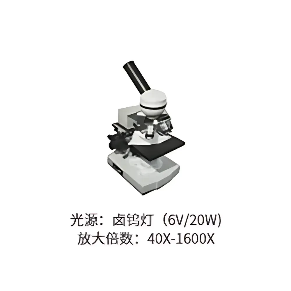
| Total Magnification Range | 40X–1600X |
| Monocular Head | 45° inclination, 360° rotation |
| Nosepiece | Four-position, outward-rolling ball bearing positioning |
| Coaxial Coarse/Fine Focus | 15 mm travel, 2 µm fine focus increment |
| Stage | Dual-layer mechanical stage, 125 × 115 mm, 70 × 30 mm movement range, 0.1 mm vernier scale |
| Mechanical Tube Length | 160 mm |
| Condenser | Abbe condenser, NA 1.25, with iris diaphragm and color filters, vertically adjustable |
| Illumination | Adjustable brightness, 6 V / 20 W halogen lamp |
| Net Weight | 3.5 kg |
| Gross Weight | 5.5 kg |
| Instrument Dimensions | 18 × 29 × 39 cm |
| Packing Dimensions | 26 × 33 × 49 cm |
Overview
The Yoke XSP-3CA Monocular Biological Microscope is a precision-engineered optical instrument designed for routine brightfield microscopy in academic teaching laboratories, clinical training environments, and quality control settings within life science and applied bioscience workflows. Built to the internationally recognized 160 mm mechanical tube length standard, it adheres to classical optical design principles that ensure compatibility with standardized eyepieces, objectives, and auxiliary components across global educational and industrial supply chains. Its optical system employs achromatic objectives corrected for chromatic aberration at two wavelengths and spherical aberration at one wavelength—providing reliable resolution and contrast for unstained and stained biological specimens including blood smears, histological sections, microbial cultures, plant tissues, and textile fibers. The monocular configuration prioritizes ergonomic simplicity and cost-effective deployment across high-density lab environments where multi-user accessibility and operational consistency are critical.
Key Features
- Optically calibrated achromatic objective set (4X, 10X, 40X spring-loaded, 100X oil immersion with spring loading) delivering consistent parfocality and minimal working distance variation across magnifications.
- Wide-field (WF) eyepieces (10X/φ18 mm and 16X/φ11 mm) engineered for extended eye relief and reduced visual fatigue during prolonged observation sessions.
- Robust coaxial coarse/fine focusing mechanism with 15 mm vertical travel and 2 µm fine focus graduation—enabling precise Z-axis localization for layered or thick specimens.
- Dual-layer mechanical stage with 70 × 30 mm travel range and 0.1 mm vernier scale for repeatable specimen positioning and comparative field-of-view documentation.
- Abbe condenser (NA 1.25) with integrated iris diaphragm and removable color filters, supporting Köhler illumination alignment and contrast optimization per ISO 8578 and ASTM E2813 guidelines.
- Stable cast-metal frame with 45° inclined, 360° rotatable monocular head—designed for long-term mechanical integrity and user-adaptive posture support.
Sample Compatibility & Compliance
The XSP-3CA supports standard 1 mm-thick glass microscope slides (76 × 26 mm) and #1.5 cover slips (0.17 mm nominal thickness), ensuring optimal performance with both dry and oil-immersion objectives. It is routinely deployed in compliance with GLP-aligned educational protocols and preliminary QC assessments governed by ISO/IEC 17025–referenced internal procedures. While not certified for diagnostic use under FDA 21 CFR Part 860 or IVD Directive 98/79/EC, its optical repeatability and mechanical stability meet baseline requirements for non-regulated morphological analysis in agricultural diagnostics, pharmaceutical excipient inspection, and undergraduate life science curricula. All halogen illumination components conform to IEC 62471 photobiological safety classifications for Class 1 LED/halogen light sources.
Software & Data Management
As a standalone optical instrument without integrated digital imaging, the XSP-3CA operates independently of proprietary software ecosystems. However, its standardized trinocular port (optional adapter available separately) enables seamless integration with third-party CMOS/CCD cameras compliant with USB 2.0/3.0 UVC protocols. When paired with open-source or commercially licensed image acquisition platforms—including ImageJ/Fiji, NIS-Elements (Nikon), or CellSens (Olympus)—the system supports timestamped image capture, basic morphometric measurement (e.g., cell diameter, mitotic index estimation), and export to TIFF/PNG formats compatible with LIMS or ELN systems. Audit trail functionality depends entirely on the external software stack; no onboard data logging or electronic records are generated natively.
Applications
- Undergraduate and graduate-level biology instruction: cell structure identification, mitosis staging, protozoan motility analysis, and fungal hyphae morphology.
- Clinical training labs: peripheral blood film review, urine sediment screening, and Gram stain interpretation under simulated diagnostic conditions.
- Agricultural extension services: seed viability assessment, nematode identification, and leaf epidermal stomatal density quantification.
- Pharmaceutical raw material testing: crystallinity evaluation of APIs, particulate contamination screening in excipients, and fiber identification in capsule shells.
- Textile QA laboratories: natural vs. synthetic fiber differentiation, weave pattern analysis, and pilling resistance assessment via surface microstructure observation.
FAQ
Is the XSP-3CA compatible with digital camera attachments?
Yes—when fitted with an optional trinocular photo-tube adapter, it supports standard C-mount or 23.2 mm eyepiece-coupled cameras.
Does the microscope include immersion oil?
No—immersion oil (Type A or B, refractive index ~1.515) must be sourced separately and is not included in the base configuration.
Can the halogen lamp be replaced with an LED module?
While not factory-supported, third-party 6 V DC LED replacements with matched beam geometry and color temperature (~6000 K) may be installed by qualified service personnel; warranty coverage does not extend to such modifications.
What maintenance intervals are recommended for routine operation?
Lens cleaning with lens tissue and reagent-grade xylene or ethanol is advised after each oil-immersion session; annual mechanical calibration of focus gearing and stage alignment is recommended for high-throughput environments.
Is the XSP-3CA suitable for fluorescence observation?
No—it lacks excitation filter sets, dichroic mirrors, and a fluorescence-capable light source; it is optimized exclusively for transmitted brightfield illumination.
Related Products


