Phenom X-view Insert for Phenom Desktop Scanning Electron Microscope
| Brand | Phenom |
|---|---|
| Country of Origin | Netherlands |
| Manufacturer Type | Original Equipment Manufacturer (OEM) |
| Origin Category | Imported |
| Model | X-view Insert |
| Pricing | Upon Request |
Overview
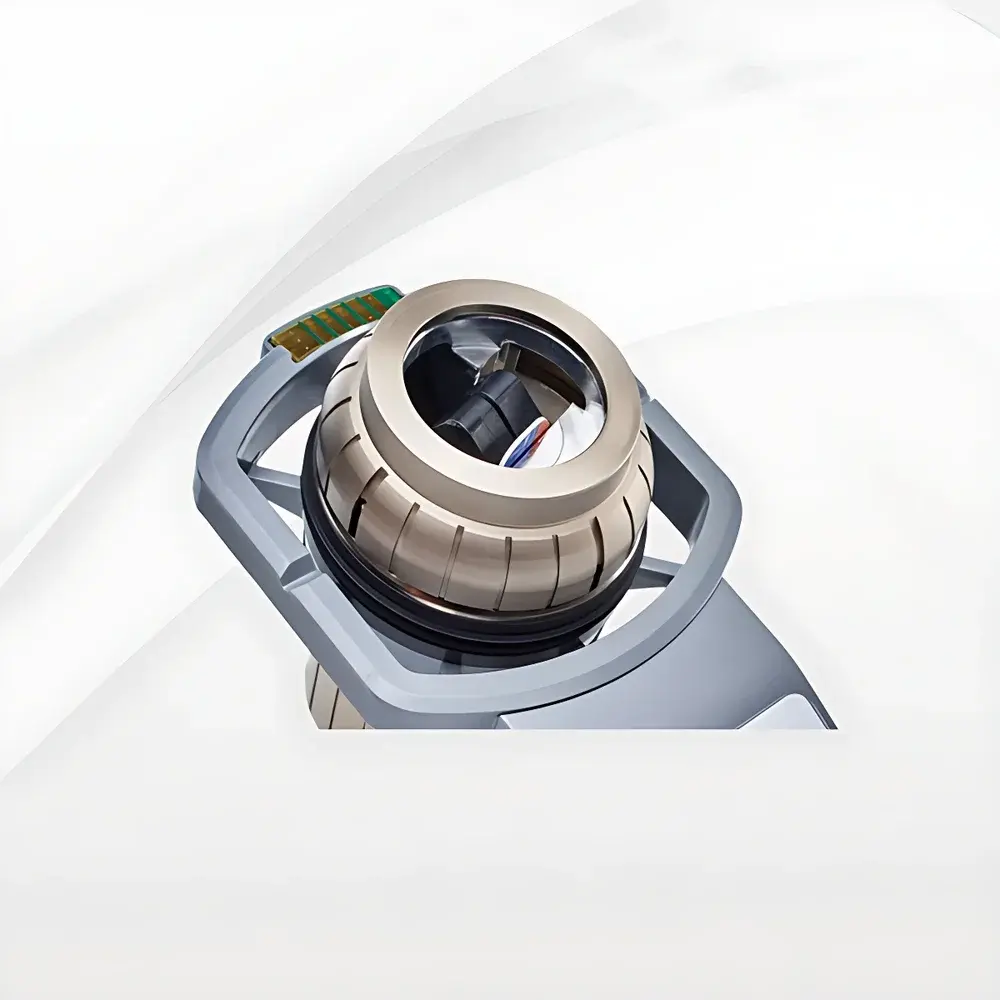
The Phenom X-view Insert is a precision-engineered mechanical sample holder accessory designed exclusively for the Phenom desktop scanning electron microscope (SEM) platform. It enables rapid, tool-free cross-sectional imaging of solid materials without requiring conventional epoxy embedding, grinding, or polishing—processes that often introduce artifacts, alter surface topography, or compromise structural integrity. Based on standard SEM imaging principles—electron beam-sample interaction generating secondary and backscattered electrons—the X-view Insert facilitates direct observation of pristine cross-sections in high vacuum or low-vacuum mode, preserving native microstructure, interfacial boundaries, coating thickness gradients, and fracture morphology. Its integration with the Phenom’s automated stage and real-time navigation system ensures sub-micron positional repeatability, making it suitable for both routine quality control and comparative failure analysis workflows.
Key Features
- Tool-free sample clamping mechanism: Eliminates reliance on screws, adhesives, or mounting resins—reducing preparation time from hours to under 60 seconds.
- Optimized geometry for unobstructed electron beam access: The insert positions samples at a fixed 45° tilt relative to the detector axis, maximizing signal collection efficiency while minimizing shadowing effects.
- Universal compatibility with Phenom standard sample cups: Designed to integrate seamlessly with the Phenom Gold, Pure, Pro, and Pharos series via the proprietary cup-mount interface.
- Robust aluminum alloy construction with electroless nickel plating: Ensures dimensional stability, electrical conductivity, and resistance to charging during extended imaging sessions.
- Adjustable lateral and rotational positioning: Fine-tuned manual alignment allows precise centering of region-of-interest (ROI) within the field of view without stage recalibration.
Sample Compatibility & Compliance
The X-view Insert accommodates flat or slightly curved specimens measuring up to 15 mm (width) × 25 mm (length) × 10 mm (thickness), including metallic alloys, polymer laminates, ceramic composites, coated substrates, and semiconductor wafers. It supports both conductive and non-conductive samples when used in conjunction with Phenom’s optional low-vacuum mode or integrated charge compensation. From a regulatory standpoint, the insert complies with ISO 14577 (instrumented indentation testing) and ASTM E1558 (SEM specimen preparation guidelines) for cross-sectional metrology. Its design aligns with GLP-compliant documentation requirements by enabling traceable, repeatable sample orientation—critical for audit-ready reports in automotive, aerospace, and medical device manufacturing environments.
Software & Data Management
When operated with Phenom’s proprietary Phenom Desktop Software (v5.5+), the X-view Insert benefits from full software integration: automatic stage coordinate mapping, user-defined ROI bookmarks, and synchronized image annotation with metadata (including insert type, sample ID, and tilt configuration). All acquired images and measurement data are stored in vendor-neutral TIFF and CSV formats, supporting interoperability with third-party analysis tools such as ImageJ, MATLAB, and Thermo Scientific Avizo. Audit trail functionality—including operator login timestamps, parameter change logs, and export history—is enabled per FDA 21 CFR Part 11 requirements when deployed in regulated laboratories.
Applications
- Coating thickness evaluation and delamination detection in automotive paint systems and PVD/CVD thin films.
- Interface characterization of multilayer dielectric stacks in MEMS and optoelectronic devices.
- Fracture surface analysis for root cause determination in metal fatigue and polymer brittle failure.
- In-process verification of solder joint integrity and wire bond cross-sections in PCB assembly lines.
- Quality assurance of encapsulated microfluidic channels and biomedical implant coatings.
FAQ
Can the X-view Insert be used with non-Phenom SEM systems?
No—it is mechanically and electronically calibrated for exclusive use with Phenom desktop SEM platforms and cannot be retrofitted to other manufacturers’ instruments.
Does the insert require grounding or additional conductive coating?
Grounding is achieved automatically through contact with the Phenom sample cup; non-conductive samples may still require carbon or gold sputtering depending on imaging conditions and magnification.
Is the X-view Insert compatible with EDS detectors?
Yes—its open geometry and minimal material occlusion allow unimpeded X-ray path transmission, enabling simultaneous high-fidelity SEM imaging and elemental mapping.
What is the maximum sample weight supported?
The insert is rated for specimens up to 200 g to maintain mechanical stability and avoid stage overload during tilt-based navigation.
How does the X-view Insert compare to traditional cross-sectioning methods in terms of measurement uncertainty?
By eliminating mechanical deformation from grinding/polishing, it reduces systematic error in layer thickness and interface position measurements by an estimated 30–50% compared to embedded sectioning, as validated in internal round-robin studies per ISO/IEC 17025 protocols.


