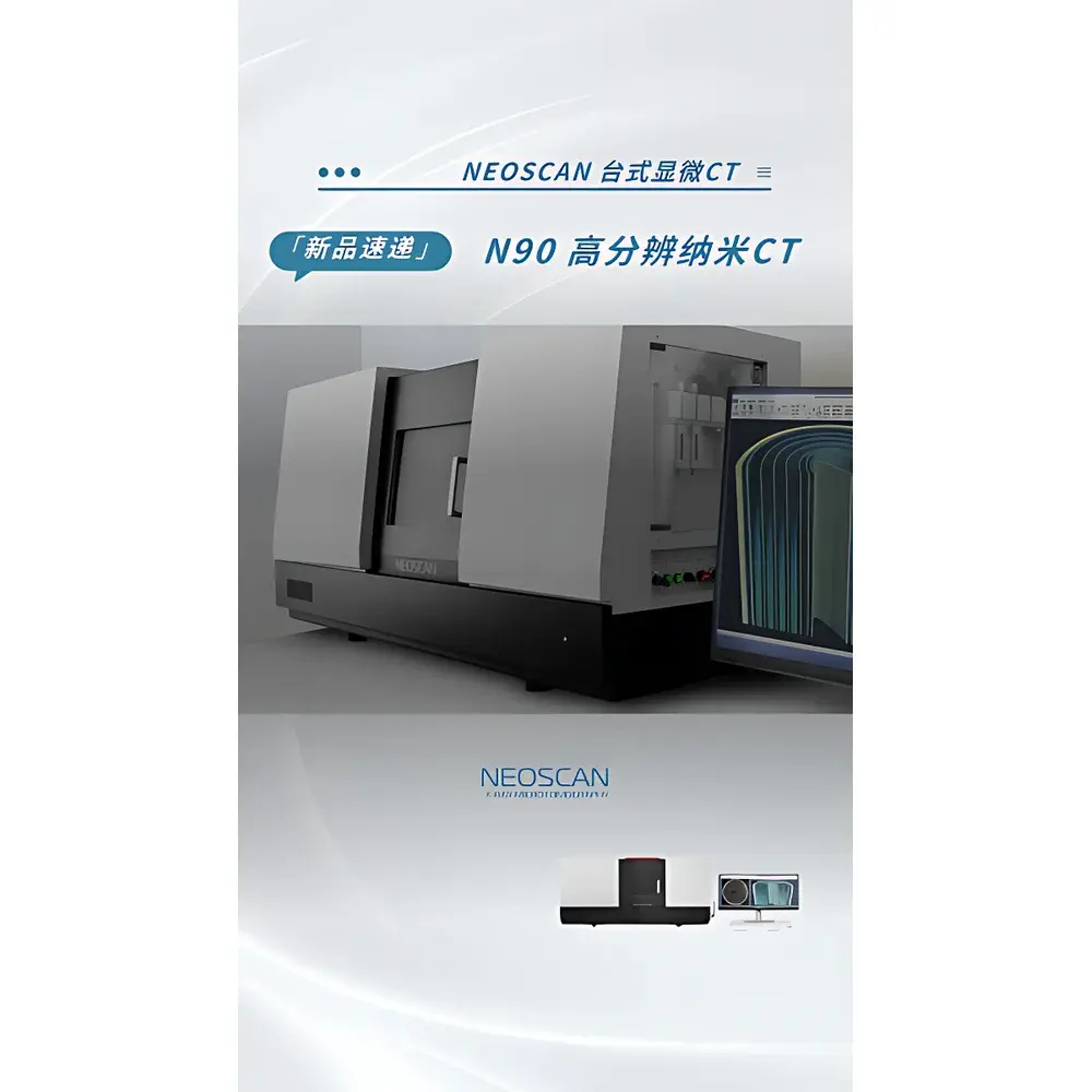
Neoscan N90 Desktop High-Resolution Nanoscale X-ray Computed Tomography System
| Brand | Neoscan |
|---|---|
| Origin | Belgium |
| Model | N90 Desktop Nanoscale CT |
| X-ray Source | 20–160 kV, 16 W Diamond-Window Microfocus Tube |
| Detector Configuration | Dual-Detector System (27 MP CMOS + 7 MP Flat-Panel Detector) |
| Spatial Resolution | ≤300 nm (≤40 nm voxel size achievable under optimized conditions) |
| Scan Mode | Rotation-Only (RO) |
| Maximum Sample Dimensions | Ø100 mm × H400 mm |
| System Dimensions | 1540 mm × 580 mm × 740 mm |
| Weight | 550 kg |
| Radiation Shielding | Integrated Lead-Lined Enclosure Compliant with IEC 61331-1 & EN 62463 |
| Filtration | Motorized 7-Position Filter Changer |
| Software Platform | NeoScan Suite v5.2 with GLP/GMP-Aware Audit Trail & FDA 21 CFR Part 11 Compliance Options |
Overview
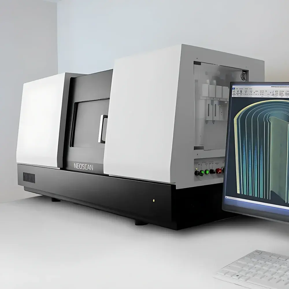
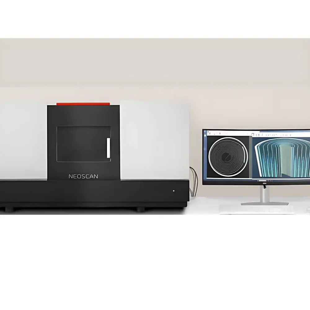
The Neoscan N90 Desktop High-Resolution Nanoscale X-ray Computed Tomography System is an engineered solution for non-destructive, three-dimensional structural characterization at sub-100 nm spatial resolution. Based on microfocus X-ray cone-beam computed tomography (CBCT), the N90 employs a high-stability diamond-window microfocus source and geometric magnification to achieve true nanoscale volumetric imaging without synchrotron dependency. Unlike conventional laboratory-scale micro-CT systems limited to micron-level resolution, the N90 delivers isotropic voxel sizes down to 40 nm—enabling quantitative analysis of pore networks in battery electrodes, intermetallic phase distribution in advanced alloys, grain boundary morphology in polycrystalline semiconductors, and ultrastructural features in mineralized biological tissues. Its rotation-only (RO) scanning geometry ensures mechanical stability and repeatability critical for time-resolved or multi-condition comparative studies. Designed for integration into standard laboratory environments—including upper-floor facilities—the N90 meets international radiation safety standards (IEC 61331-1, EN 62463) and operates within Class II X-ray equipment regulatory frameworks.
Key Features
- Desktop form factor (1540 × 580 × 740 mm; 550 kg) enables deployment in ISO Class 5–7 cleanrooms, university core labs, and industrial R&D spaces without structural reinforcement.
- Dual-detector architecture: A 27 MP scientific CMOS detector (16-bit dynamic range, <0.5 e⁻ read noise) for high-fidelity nanoscale imaging; and a 7 MP flat-panel detector for rapid large-field-of-view surveys and coarse-to-fine workflow bridging.
- 20–160 kV, 16 W diamond-window microfocus X-ray source with active thermal management and <1 µm focal spot stability over 10,000-hour lifetime—optimized for low-Z to high-Z material contrast across Li-ion cathodes, SiC wafers, and calcified bone.
- Motorized 7-position filter changer with automatic selection logic integrated into acquisition protocols—reducing beam hardening artifacts and enabling consistent quantitative attenuation calibration per sample class.
- Integrated oil-free vacuum system and pre-aligned filament cartridge simplify preventive maintenance and reduce mean time to repair (MTTR) by >65% versus legacy systems requiring manual source realignment.
- Real-time remote diagnostics with SNMP-based telemetry monitoring of tube voltage/current, detector temperature, enclosure interlock status, and cooling fan RPM—supporting predictive maintenance and GLP-compliant service logging.
Sample Compatibility & Compliance
The N90 accommodates cylindrical samples up to Ø100 mm × 400 mm height and supports both rigid and semi-rigid specimens—including packaged semiconductor dies, sintered ceramic pellets, freeze-dried tissue biopsies, and encapsulated electrochemical cells. Sample mounting utilizes standardized kinematic fixtures compatible with ASTM E1441-22 (Standard Guide for Computed Tomography) and ISO/IEC 17025-accredited workflows. Radiation shielding complies with EU Directive 2013/59/Euratom and U.S. NRC 10 CFR Part 20 requirements. Optional XRF module (not included by default) enables elemental mapping co-registered with CT volume data per ASTM E1508-21 (Standard Practice for Quantitative Analysis by Energy Dispersive Spectrometry). System software supports audit trail generation, electronic signatures, and role-based access control aligned with FDA 21 CFR Part 11 and EU Annex 11 expectations.
Software & Data Management
NeoScan Suite v5.2 provides end-to-end workflow automation—from geometry calibration and projection acquisition to iterative reconstruction (SART, SIRT), phase retrieval enhancement, and quantitative morphometry. Reconstruction engine supports GPU-accelerated FDK and model-based algorithms with regularization tuned for low-signal-to-noise nanoscale datasets. All processing steps are scriptable via Python API (PyNeoScan), enabling integration into automated analysis pipelines compliant with FAIR data principles. Project metadata—including acquisition parameters, calibration certificates, and operator credentials—is embedded in DICOM-CT and NIfTI-1 containers. Raw projections and reconstructed volumes are stored in vendor-neutral HDF5 format with SHA-256 checksums for integrity verification. Long-term archival complies with ISO 16363:2012 (Trusted Digital Repository criteria).
Applications
- Lithium-ion battery R&D: Quantification of SEI thickness heterogeneity, cathode particle cracking, and pore tortuosity evolution during cycling—correlated with electrochemical impedance spectroscopy (EIS) data.
- Semiconductor packaging: Detection of voids, delamination, and Cu diffusion in TSV (through-silicon via) structures at <100 nm scale, supporting JEDEC JESD22-B111 failure analysis protocols.
- Advanced materials: 3D grain reconstruction in additively manufactured Inconel 718, segmentation of second-phase precipitates in Al-Mg-Si alloys, and fatigue crack initiation mapping in Ti-6Al-4V.
- Life sciences: Mineral density distribution in osteocyte lacunocanalicular networks, collagen fibril orientation in tendon cross-sections, and vascular perfusion modeling in decellularized lung scaffolds.
- Geosciences: Pore-throat network extraction from shale matrix samples, clay mineral distribution analysis in reservoir rock cores, and fossilized microstructure preservation assessment.
FAQ
What is the minimum achievable voxel size under routine operating conditions?
The N90 achieves isotropic voxels of 40 nm in optimized configurations (e.g., small field-of-view, high magnification, extended exposure), validated using NIST-traceable line-pair test objects (NIST SRM 2058). Typical operational voxel sizes range from 60–300 nm depending on sample size and required signal-to-noise ratio.
Does the system support in situ or operando experiments?
Yes—mechanical stages with ±10 µm positioning repeatability and programmable thermal chambers (–20 °C to +150 °C) are available as factory-integrated options. Electrical feedthroughs support simultaneous electrochemical testing during tomographic acquisition.
How is radiation safety ensured during daily operation?
The fully enclosed lead-lined cabinet incorporates dual redundant door interlocks, real-time dose rate monitoring (SiPM-based), and automatic beam shutdown upon interlock breach—meeting Type B radiation device classification per IEC 61331-1.
Is the NeoSpace 3D visualization module included by default?
No—the NeoSpace裸眼 3D module is an optional hardware add-on featuring a 27″ autostereoscopic display with eye-tracking-enabled parallax correction and native support for DICOM-CT volume rendering without external VR infrastructure.
Can raw projection data be exported for third-party reconstruction?
Yes—raw TIFF stacks (16-bit unsigned integer) and metadata XML files are exportable via USB 3.2 Gen 2 or GigE interface. Calibration files (flat/dark field, geometry matrices) follow the X-ray CT Metadata Standard (XCT-MS) v1.1 specification.


