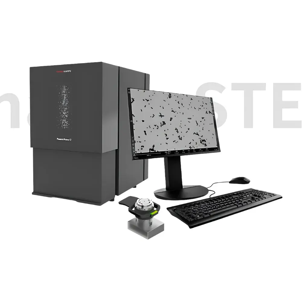
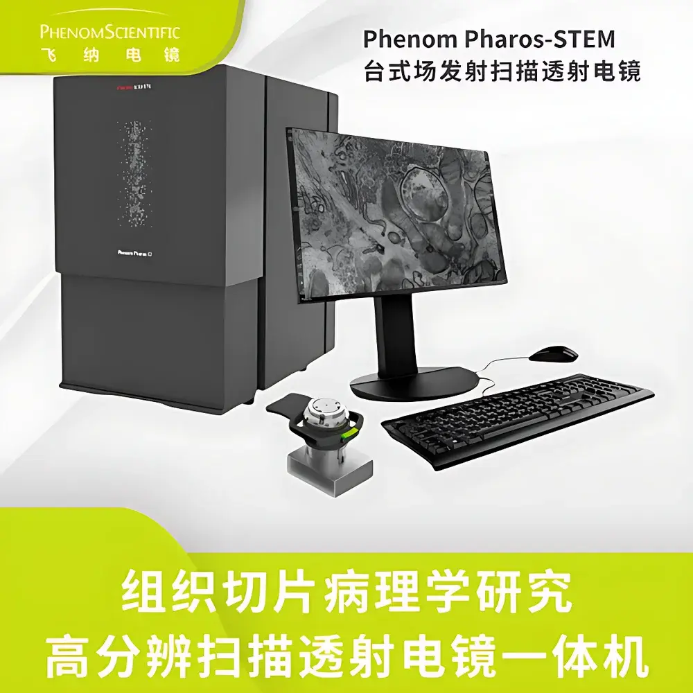
Phenom Pharos STEM Desktop Field-Emission Scanning Transmission Electron Microscope
| Brand | Phenom |
|---|---|
| Origin | Netherlands |
| Manufacturer Type | Original Equipment Manufacturer (OEM) |
| Origin Category | Imported |
| Model | Pharos STEM |
| Pricing | Available Upon Request |
Overview
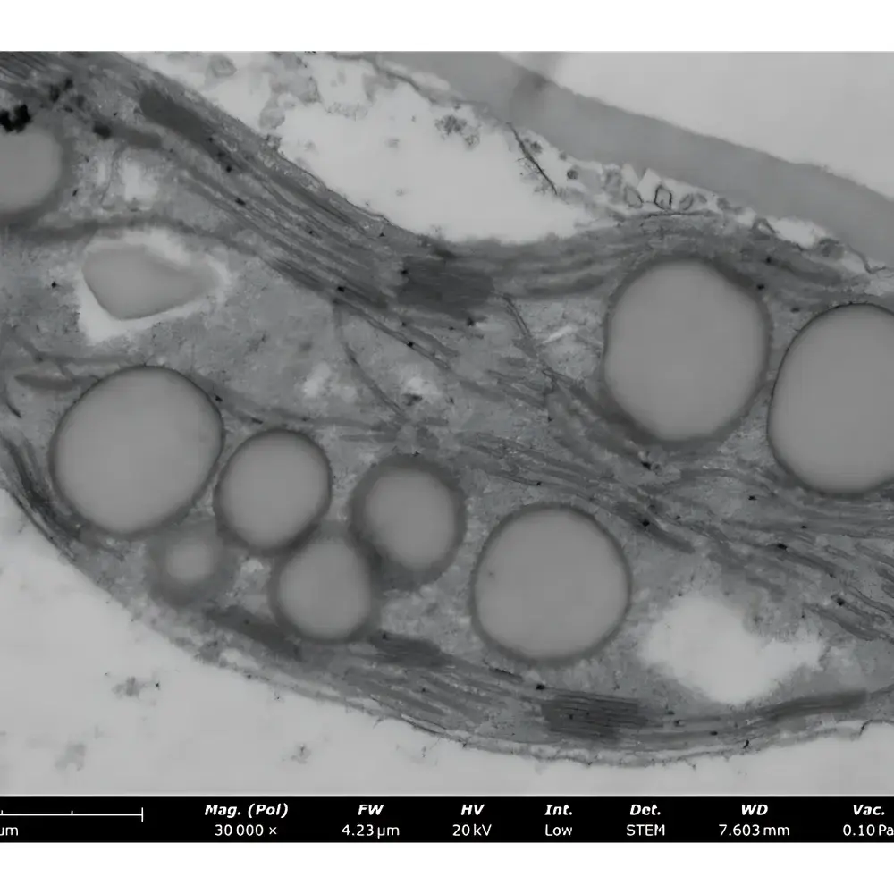
The Phenom Pharos STEM is a desktop field-emission scanning transmission electron microscope engineered for high-resolution structural and compositional analysis of ultrathin biological and material specimens. Unlike conventional TEMs requiring dedicated high-vacuum rooms and complex sample preparation, the Pharos STEM integrates STEM functionality into a compact, user-friendly benchtop platform. It operates on the principle of scanning transmission electron microscopy (STEM), where a focused electron probe raster-scans across a thin specimen (<100 nm), and transmitted/scattered electrons are collected by segmented solid-state detectors to generate bright-field (BF), dark-field (DF), and high-angle annular dark-field (HAADF) images in parallel. Its field-emission gun (FEG) delivers stable, high-brightness electron beams across an accelerating voltage range of 1–20 kV—enabling optimal contrast control while minimizing beam-induced damage to delicate biological tissues such as formalin-fixed paraffin-embedded (FFPE) or cryo-ultrathin sections used in histopathological research.
Key Features
- Desktop-integrated STEM capability—world’s only commercially available benchtop STEM system compatible with the Phenom Pharos G2 platform
- FEG electron source ensuring sub-nanometer resolution (≤1 nm at 20 kV) and exceptional signal-to-noise ratio across low-to-medium kV operation
- Dedicated STEM sample holder accommodating standard 3 mm TEM grids with mechanical clamping—no specialized grid manipulation tools required
- 11-segment annular detector array enabling fully customizable imaging: simultaneous acquisition of BF, DF, and HAADF signals with real-time detector weighting
- Fixed working distance and automated detector alignment—eliminating manual optimization and reducing operator dependency
- Full UI integration within Phenom’s proprietary software suite, supporting one-click STEM workflow initiation and parameter recall
- Imaging time ≤40 seconds from sample loading to final image display—optimized for rapid screening and iterative experimental design
Sample Compatibility & Compliance
The Pharos STEM is validated for use with routinely prepared histopathological specimens, including resin-embedded tissue sections (e.g., epoxy or acrylic), carbon-coated ultrathin sections (50–80 nm), and unstained or lightly stained (e.g., uranyl acetate/lead citrate) TEM grids. Its low-kV STEM mode (1–5 kV) significantly reduces radiolysis and mass loss in organic matrices—critical for preserving antigenicity and ultrastructural fidelity in diagnostic pathology workflows. The system complies with ISO 14644-1 Class 5 cleanroom requirements for internal chamber environment and meets CE marking standards for electromagnetic compatibility (EN 61326-1) and safety (EN 61010-1). While not FDA-cleared as a diagnostic device, its operational parameters align with GLP-compliant documentation practices, including audit-trail-enabled session logging and timestamped image metadata export per ISO/IEC 17025 guidelines.
Software & Data Management
Phenom’s proprietary software provides a unified interface for SEM and STEM modalities, featuring intuitive drag-and-drop STEM detector configuration, real-time histogram-based contrast adjustment, and batch-mode image export in TIFF, PNG, and DM3 formats. All acquired STEM datasets retain full detector segmentation metadata, enabling post-acquisition reweighting of BF/DF/HAADF contributions without rescanning. Image metadata includes vacuum pressure logs (0.1–60 Pa range), beam current values, dwell time, pixel dwell, and stage coordinates—supporting traceability in regulated environments. Raw data files are structured for interoperability with third-party analysis platforms (e.g., DigitalMicrograph, ImageJ/Fiji) and support batch processing via Python API hooks for automated feature extraction and classification pipelines.
Applications
- Subcellular ultrastructure analysis in renal glomeruli, pancreatic acinar cells, and neural synapses—correlating light microscopy diagnoses with nanoscale morphology
- High-contrast visualization of low-Z biological macromolecules (e.g., viral capsids, collagen fibrils, lipid droplets) using BF mode
- Z-contrast mapping of heavy-metal-stained organelles (e.g., mitochondria, lysosomes) and mineral deposits (e.g., calcium phosphate) via HAADF imaging
- Crystallographic contrast enhancement in amyloid fibrils and prion aggregates using DF mode for diffraction-sensitive contrast
- Rapid quality assessment of section thickness uniformity and staining homogeneity prior to high-throughput TEM tomography
- Materials science applications including nanoparticle dispersion analysis, catalyst support interactions, and polymer phase segregation
FAQ
Is the Pharos STEM compatible with standard TEM grids?
Yes—it accepts standard 3 mm diameter copper, nickel, or gold TEM grids secured via a precision-machined mechanical clamp; no grid welding or electrostatic mounting is required.
Can BF, DF, and HAADF images be acquired simultaneously?
Yes—the 11-segment detector enables concurrent signal acquisition across all three modes, with real-time weighted summation configurable via the GUI.
What vacuum levels does the STEM column operate at during imaging?
The system maintains stable imaging conditions at three preset vacuum levels: 0.1 Pa (high-resolution STEM), 10 Pa (fast survey mode), and 60 Pa (rapid loading/unloading); all transitions are automated and pressure-stabilized.
Does the Pharos STEM require liquid nitrogen or external water cooling?
No—it employs air-cooled FEG and solid-state detectors, eliminating cryogenic dependencies and enabling continuous 24/7 operation in standard laboratory environments.
How is image calibration performed?
Calibration is performed automatically using built-in grating standards and verified via NIST-traceable reference samples; scale bars are embedded in all exported images with uncertainty reporting.


