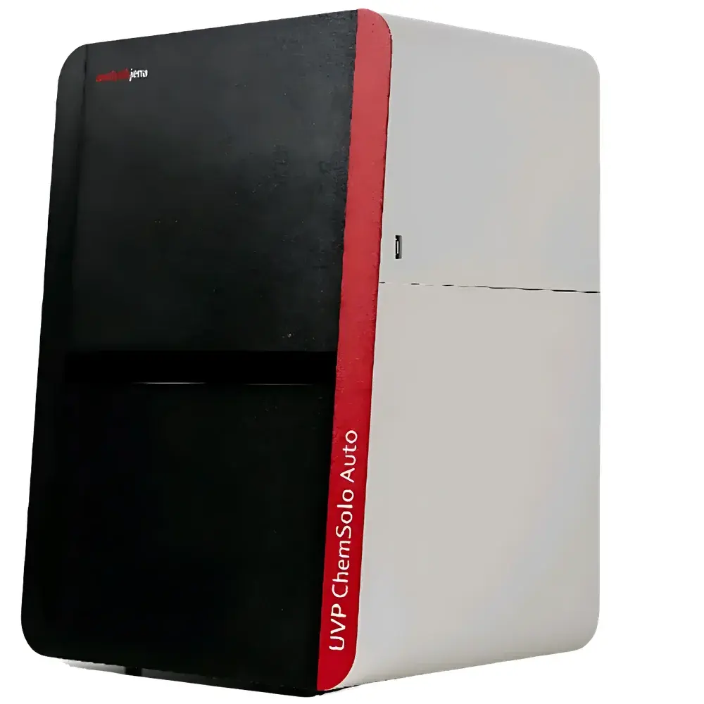
UVP ChemSolo Auto Chemiluminescence Imaging System
| Brand | Analytik Jena |
|---|---|
| Origin | USA |
| Manufacturer | Analytik Jena GmbH |
| Country of Origin | Imported |
| Model | UVP ChemSolo Auto |
| Pixel Size | 3.76 µm × 3.76 µm |
| Sensor Dimensions | 11.3 mm × 11.3 mm |
| Dynamic Range | 4.8 OD |
| Quantum Efficiency | 85% |
| Read Noise | 1.5 e⁻ |
| Dark Current | 0.0005 e⁻/p/s |
| Sensor Type | sCMOS (9 MP) |
| Binning Options | 1×1 to 8×8 |
| Compatible Light Sources | Long-life cold-cathode UV transilluminator (20-year lifespan) |
| Supported Image Formats | JPEG, BMP, TIFF |
| Applications | Nucleic Acid Imaging, Protein Gel Imaging, Chemiluminescent Western Blot Detection |
Overview
The UVP ChemSolo Auto Chemiluminescence Imaging System is a high-performance, fully integrated optical imaging platform engineered for quantitative molecular biology applications in academic research laboratories, pharmaceutical QC facilities, and contract research organizations (CROs). Developed by Analytik Jena—formed through the strategic integration of the legacy UVP (Ultra-Violet Products) imaging expertise and Zeiss-derived optical engineering heritage—the system leverages a scientific-grade back-illuminated sCMOS sensor to deliver exceptional sensitivity and dynamic fidelity across multiple detection modalities. Unlike conventional CCD-based systems, the ChemSolo Auto employs a large-format 9-megapixel sCMOS detector with 3.76 µm pixel pitch and 11.3 mm × 11.3 mm active area, enabling high-resolution spatial sampling without compromising field-of-view coverage. Its core measurement principle relies on photon capture from chemiluminescent, fluorescent, and UV-excited emissions, with signal acquisition governed by precise exposure control, temperature-stabilized sensor operation, and calibrated optical path geometry. The system is purpose-built for reproducible quantification of Western blots, nucleic acid gels, and protein expression assays under both ambient and darkroom conditions.
Key Features
- Back-illuminated sCMOS sensor with 85% peak quantum efficiency and 4.8 optical density (OD) dynamic range for accurate multi-band signal capture
- Ultra-low read noise (1.5 e⁻ RMS) and sub-pixel dark current (0.0005 e⁻/p/s), ensuring high signal-to-noise ratio even at short exposure times
- Integrated long-life cold-cathode UV transilluminator with >20,000-hour operational lifetime and minimal thermal output—reducing gel distortion and eliminating frequent lamp replacement
- Flexible binning architecture (1×1 to 8×8) to optimize resolution versus sensitivity trade-offs across diverse sample types and emission intensities
- Dedicated chemiluminescence imaging tray with optimized reflectivity and background suppression geometry
- Modular accessory ecosystem including gel transfer铲 (with drainage apertures), physiological temperature-controlled animal imaging plate, and UV-safe viewing shield
Sample Compatibility & Compliance
The ChemSolo Auto supports standard electrophoretic formats including mini-gels (up to 15 cm × 15 cm), SDS-PAGE, agarose, and native PAGE gels, as well as membrane-based chemiluminescent blots (PVDF and nitrocellulose). It complies with ISO/IEC 17025 requirements for laboratory equipment qualification when used in validated workflows. While not inherently 21 CFR Part 11 compliant out-of-the-box, the open software architecture enables integration with third-party electronic lab notebook (ELN) and LIMS platforms supporting audit trail, user authentication, and data integrity controls. The system meets IEC 61000-6-3 (EMC) and IEC 61000-6-2 (immunity) standards and is CE-marked for use in EU laboratories. All optical components—including Zeiss-designed relay lenses and bandpass filters—are traceably calibrated per manufacturer specifications and documented in the instrument’s factory certificate of conformance.
Software & Data Management
The ChemSolo Auto ships with UVP VisionWorks LS software—a dual-open platform that supports unrestricted installation on Windows-based workstations and natively reads TIFF, JPEG, and BMP files from external sources (e.g., scanners, microscopes, or legacy imaging systems). The software provides full quantitative densitometry with background subtraction, lane/band identification, molecular weight calibration, and relative intensity normalization. Advanced modules support colony counting (CFU analysis), plant phenotyping (leaf area, chlorophyll index), and small-animal bioluminescence/chemiluminescence imaging with ROI-based kinetic profiling. Raw image metadata—including exposure time, gain setting, binning mode, lens aperture, and sensor temperature—is embedded in EXIF-compliant headers. Export options include CSV for statistical analysis, PDF reports with embedded annotations, and PNG/TIFF with 16-bit linear scaling for downstream processing in ImageJ/Fiji or MATLAB.
Applications
- Quantitative Western blot analysis using HRP- or AP-conjugated secondary antibodies with ECL or CDP-Star substrates
- Nucleic acid visualization via ethidium bromide, SYBR Safe, or GelRed staining under 302 nm UV excitation
- Protein gel documentation with Coomassie, silver, or fluorescent dyes (e.g., Deep Purple)
- Low-light bioluminescent imaging of luciferase-expressing cell lines or transgenic models
- High-throughput colony screening and morphological classification in microbiology
- Plant root/shoot imaging with chlorophyll fluorescence or histochemical staining (e.g., GUS)
FAQ
Does the ChemSolo Auto support time-lapse chemiluminescence imaging?
Yes—software-defined exposure sequences with programmable intervals enable kinetic acquisition over durations up to 24 hours.
Can the system be validated for GLP/GMP environments?
Yes—through documented IQ/OQ protocols, user-accessible calibration logs, and optional 21 CFR Part 11 add-ons provided by certified third-party vendors.
Is the UV transilluminator compatible with ethidium bromide alternatives such as SYBR Gold?
Yes—the 302 nm cold-cathode source delivers optimal excitation efficiency for SYBR Gold, GelGreen, and other modern nucleic acid stains.
What is the maximum supported file size for batch analysis?
The software handles stacks of up to 500 TIFF images (16-bit, 9 MP) in memory with real-time preview and parallel processing.
Are lens and filter specifications published for optical validation?
Yes—full optical schematics, MTF curves, and spectral transmission data are included in the technical appendix of the user manual and available upon request from Analytik Jena Technical Support.


