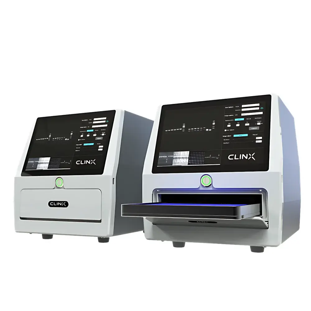
CLINX GenoSens S2 Mini All-in-One Gel Imaging System
| Brand | CLINX |
|---|---|
| Origin | Shanghai, China |
| Model | GenoSens S2 |
| Instrument Type | Standard Gel Imaging System |
| CCD Resolution | 6.37 MP (3089 × 2064 pixels) |
| Bit Depth | 16-bit |
| Lens | High-Resolution Fixed-Focal Lens |
| Imaging Area | 16 × 11.5 cm |
| UV Transillumination | 302 nm LED (17 × 13 cm active area) |
| Blue Light Transillumination | 470 nm LED (17 × 13 cm) |
| White Light Reflectance | Integrated LED |
| Filter | Multi-layer 99% UV-blocking emission filter |
| Sample Stage | Motorized adjustable stage with auto-alignment |
| Interface | USB 2.0, Wi-Fi module optional |
| Touchscreen | Embedded 10.1-inch capacitive multi-touch display |
| Dimensions (W×D×H) | 386 × 355 × 321 mm |
| Compliance | CE, RoHS, ISO 13485–compatible design for laboratory use |
Overview
The CLINX GenoSens S2 Mini All-in-One Gel Imaging System is an integrated, benchtop gel documentation platform engineered for precision, reproducibility, and workflow efficiency in molecular biology laboratories. Built upon a solid-state LED transillumination architecture and a high-sensitivity 16-bit monochrome CCD sensor, the system captures fluorescence and visible-light images of nucleic acid and protein gels via controlled excitation and selective emission filtering. Its optical path incorporates a fixed-focal, low-distortion lens optimized for uniform field illumination and minimal vignetting across the full 16 × 11.5 cm imaging area. Unlike modular systems requiring external computers or manual alignment, the GenoSens S2 Mini integrates acquisition hardware, motorized stage control, and real-time image processing into a single compact enclosure—reducing footprint, eliminating cable clutter, and minimizing inter-unit calibration drift. The system operates under ambient lab lighting without requiring darkroom conditions, supporting routine QC, teaching labs, and regulated environments where traceability and operator independence are essential.
Key Features
- 6.37-megapixel monochrome CCD sensor with true 16-bit dynamic range (65,536 intensity levels), enabling quantification across broad signal intensities without saturation or noise floor limitations.
- Motorized sample stage with automatic height and position adjustment—synchronized with selected light source (UV 302 nm, blue 470 nm, or white reflectance) to maintain optimal focus and illumination uniformity.
- Dual-wavelength LED transillumination: 302 nm UV for ethidium bromide and SYBR dyes; 470 nm blue light for safer nucleic acid visualization with SYBR Safe, GelRed, or GelGreen—reducing photodamage and mutagenic risk.
- Integrated 10.1-inch multi-touch LCD interface with embedded GS2Mini acquisition software—enabling fully autonomous operation without external PC dependency.
- Intelligent gel tray recognition: automatically detects tray type (e.g., standard minigel, midi-gel, or custom cassette) and selects appropriate exposure parameters, lens focus, and background subtraction profile.
- Wi-Fi connectivity option for wireless image export, remote monitoring, and direct printing—compatible with standard IEEE 802.11n infrastructure and TLS-secured data transmission.
- UV-blocking emission filter with >99% attenuation at <300 nm, meeting IEC 62471 photobiological safety requirements for Class 1 LED devices.
Sample Compatibility & Compliance
The GenoSens S2 Mini accommodates standard vertical and horizontal electrophoresis gels—including agarose (0.5–4%), polyacrylamide (8–20%), and gradient gels—up to 16 × 11.5 cm. It supports common nucleic acid stains (Ethidium Bromide, SYBR Green I/II, SYBR Safe, GelRed, GelGreen, GelStar) and protein stains (Coomassie Brilliant Blue R-250/G-250, Silver Nitrate, SYPRO Ruby, SYPRO Orange). For regulatory compliance, the system’s firmware logs all acquisition parameters (exposure time, gain, lens position, light source, filter state) with timestamped audit trails—supporting GLP and GMP-aligned workflows. While not FDA 21 CFR Part 11 certified out-of-box, its software architecture permits integration with validated LIMS or ELN platforms through standardized DICOM or TIFF export protocols. All electrical components conform to CE marking directives (2014/30/EU EMC, 2014/35/EU LVD) and RoHS 2011/65/EU material restrictions.
Software & Data Management
The embedded GS2Mini acquisition software provides guided, context-aware imaging: users select application mode (DNA gel, protein gel, or white-light documentation), and the system calculates optimal exposure using real-time histogram analysis—not preset timers. Image capture includes auto-bracketing, region-of-interest (ROI) masking, and live contrast preview. Post-acquisition, the optional PC-based GelAnalysis Pro software enables quantitative densitometry compliant with ISO/IEC 17025 principles: background subtraction (rolling ball or polynomial fit), lane and band detection (adaptive thresholding), molecular weight calibration (log-linear regression against ladder standards), and relative quantification (normalized band intensity vs. reference control). All results export to CSV or Excel-compatible XLSX formats; TIFF files embed EXIF metadata including exposure settings, camera temperature, and user ID. Audit trail reports record every software action—parameter change, image save, analysis run—with SHA-256 hash integrity verification.
Applications
- Nucleic acid gel documentation: high-fidelity imaging of PCR products, restriction digests, and sequencing ladders stained with low-toxicity dyes—optimized for downstream applications such as band excision and cloning.
- Protein electrophoresis analysis: sensitive detection of Coomassie- and silver-stained SDS-PAGE gels, including post-translational modification profiling when paired with fluorescent dyes like SYPRO Ruby.
- Gel slice excision support: integrated UV shielding hood (optional accessory) meets EN 60825-1 laser product safety guidelines during manual band cutting under 302 nm illumination.
- Teaching and core facility use: intuitive touch interface lowers training burden; consistent output reduces inter-operator variability in undergraduate labs or shared resource centers.
- Longitudinal study documentation: time-series gel imaging with automated file naming, version-controlled metadata, and batch processing ensures traceability across multi-week experiments.
FAQ
Does the GenoSens S2 Mini require a dedicated computer for operation?
No—the system runs natively on its embedded ARM-based controller with touchscreen UI. A PC is only required for advanced analysis using GelAnalysis Pro or for archival transfer via USB/Wi-Fi.
Can it image SYBR Gold-stained gels under blue light?
Yes—its 470 nm blue LED excites SYBR Gold efficiently, delivering signal-to-noise ratios comparable to UV excitation while significantly reducing DNA damage and operator UV exposure.
Is the CCD cooled, and what is its dark current specification?
The sensor operates at ambient temperature with thermally stabilized analog front-end circuitry; typical dark current is ≤0.005 e⁻/pixel/sec at 25°C—sufficient for exposures up to 60 seconds without active cooling.
How does the motorized stage ensure repeatable positioning across multiple users?
Each stage movement is encoder-referenced and calibrated during startup; positional offsets are stored per user profile and persist across power cycles, ensuring inter-user reproducibility within ±0.1 mm.
Are raw image files saved in an open format suitable for third-party analysis?
Yes—TIFF files are saved with uncompressed 16-bit grayscale data, EXIF metadata, and no proprietary compression; compatible with ImageJ/Fiji, MATLAB, Python (OpenCV/scikit-image), and commercial bioinformatics suites.
Related Products


