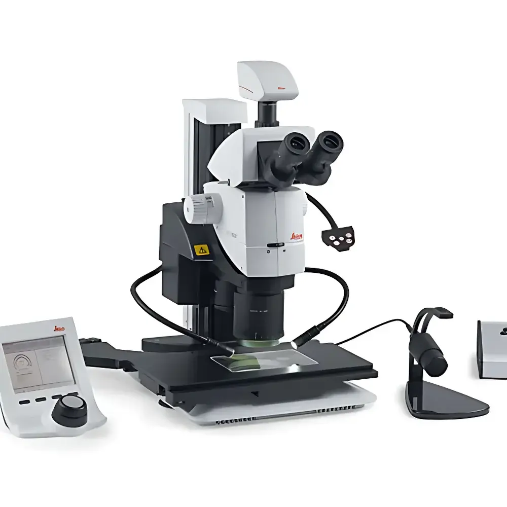
Leica M125 C & M205 C Encoded Stereo Microscopes
| Brand | Leica |
|---|---|
| Origin | Germany |
| Model | Leica M125 C & M205 C |
| Total Magnification | 800× |
| Zoom Ratio | 12.5:1 (M125 C), 20.5:1 (M205 C) |
| Field Diameter | ∅ 68 mm |
| Working Distance | 61.5 mm |
| Objective Type | 2.0× PlanApo |
| Optical System | 100% Apochromatic Correction |
| Numerical Aperture | 0.288 |
| Resolution | 864 lp/mm (M125 C, with 2.0× objective), 1050 lp/mm (M205 C, with 2.0× objective) |
| Minimum Resolvable Feature Size | 0.952 µm (M205 C) |
Overview
The Leica M125 C and M205 C are high-performance encoded stereo microscopes engineered for precision inspection, quality control, and research applications in regulated and demanding laboratory environments. Both models operate on the principle of Greenough optical design—dual independent optical paths that deliver true stereoscopic vision—and integrate Leica’s proprietary FusionOptics technology. This dual-path architecture simultaneously optimizes resolution in one channel and depth of field in the other; the human visual system fuses these complementary image streams into a single, high-fidelity 3D representation with exceptional sharpness, contrast, and spatial fidelity. Unlike conventional stereo microscopes constrained by the inverse relationship between resolution and depth of field, FusionOptics decouples these parameters, enabling consistent visualization of fine surface topography, sub-micron structural features, and complex layered samples without compromise. The M125 C delivers up to 864 line pairs per millimeter resolution with its 2.0× PlanApo objective, while the M205 C achieves 1050 lp/mm and resolves features down to 0.952 µm—making it the highest-resolution optical stereo microscope commercially available at the time of its introduction.
Key Features
- Encoded zoom and focus mechanisms for automated parameter capture and reproducible setup recall
- FusionOptics technology: real-time parallel optimization of resolution and depth of field across dual optical paths
- 100% apochromatically corrected optical train ensuring color fidelity, minimal chromatic aberration, and high transmission efficiency across visible wavelengths
- Integrated dual variable aperture diaphragms for dynamic control of contrast, depth of field, and illumination uniformity
- Ergonomic modular design compliant with ISO 9241-5 and EN 614-1 standards for sustained operator comfort during extended inspection sessions
- PlanApo 2.0× objective with numerical aperture of 0.288 and working distance of 61.5 mm—optimized for high-resolution surface metrology and non-contact dimensional assessment
- Field diameter of ∅ 68 mm supports large-area sample evaluation without stage repositioning
Sample Compatibility & Compliance
The M125 C and M205 C accommodate a broad range of opaque, translucent, and reflective specimens—including polished wafers, solder joints, medical device components, polymer composites, and biological tissue sections—without requiring conductive coating or vacuum environments. Their long working distance and large field of view support integration with motorized XY stages, micromanipulators, and environmental chambers. Both systems comply with IEC 61000-6-3 (EMC emissions), IEC 61000-6-2 (immunity), and meet CE marking requirements for laboratory equipment. When paired with Leica Application Suite X (LAS X) software, they support audit trails, electronic signatures, and metadata embedding aligned with FDA 21 CFR Part 11 and EU Annex 11 for regulated QC/QA workflows. Calibration certificates traceable to PTB (Physikalisch-Technische Bundesanstalt) are available upon request.
Software & Data Management
Leica LAS X provides full bidirectional communication with encoded hardware: magnification, aperture position, focus height, and illumination intensity are automatically logged with every acquired image. Scale bars are dynamically overlaid and recalculated in real time during zoom changes. Image metadata—including objective ID, exposure settings, calibration status, and instrument serial number—is embedded in TIFF and JPEG files using EXIF and DICOM-SR compliant tags. Batch acquisition, multi-focus stacking, and measurement annotation tools enable standardized reporting across shifts and sites. All configuration presets can be exported as XML profiles for deployment across multiple instruments—ensuring inter-laboratory consistency and simplifying GLP/GMP documentation.
Applications
- Semiconductor packaging inspection: bond wire integrity, die attach void detection, lead frame coplanarity assessment
- Medical device manufacturing: catheter tip inspection, stent strut analysis, suture knot verification under ISO 13485-compliant processes
- Materials science: grain boundary mapping in metallurgical samples, fiber orientation in composites, fracture surface morphology
- Electronics assembly: solder joint wetting angle quantification, BGA void fraction analysis, conformal coating thickness estimation
- Forensic document examination: ink differentiation, paper fiber structure, latent impression enhancement
- Academic research: developmental biology specimen staging, entomological morphology, paleontological fossil surface reconstruction
FAQ
What distinguishes FusionOptics from conventional stereo microscopy?
FusionOptics employs two physically separate optical paths—one optimized for maximum resolution, the other for maximum depth of field—whose outputs are fused perceptually by the observer. This eliminates the traditional trade-off between detail clarity and volumetric context.
Are the M125 C and M205 C compatible with third-party digital cameras?
Yes—both models feature standardized C-mount and F-mount adapters, supporting integration with scientific CMOS/CCD cameras from vendors including Basler, IDS, and FLIR, provided appropriate driver support is available in LAS X.
Can encoded settings be synchronized across multiple instruments in a production lab?
Yes—LAS X allows centralized profile management via networked license servers, enabling identical calibration states and imaging protocols across distributed Leica stereo platforms.
Is routine calibration required for quantitative measurements?
While optical alignment remains stable due to monolithic mechanical construction, annual verification against NIST-traceable stage micrometers is recommended for ISO/IEC 17025-accredited laboratories performing dimensional metrology.
Do these microscopes support fluorescence observation?
No—neither model includes epi-illumination pathways or filter cube turrets; they are designed exclusively for reflected and transmitted brightfield, oblique, and polarized light illumination.


