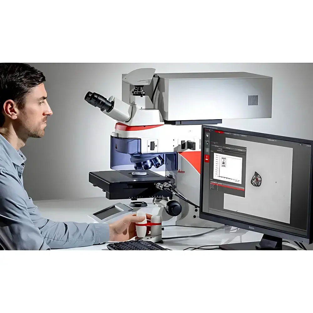
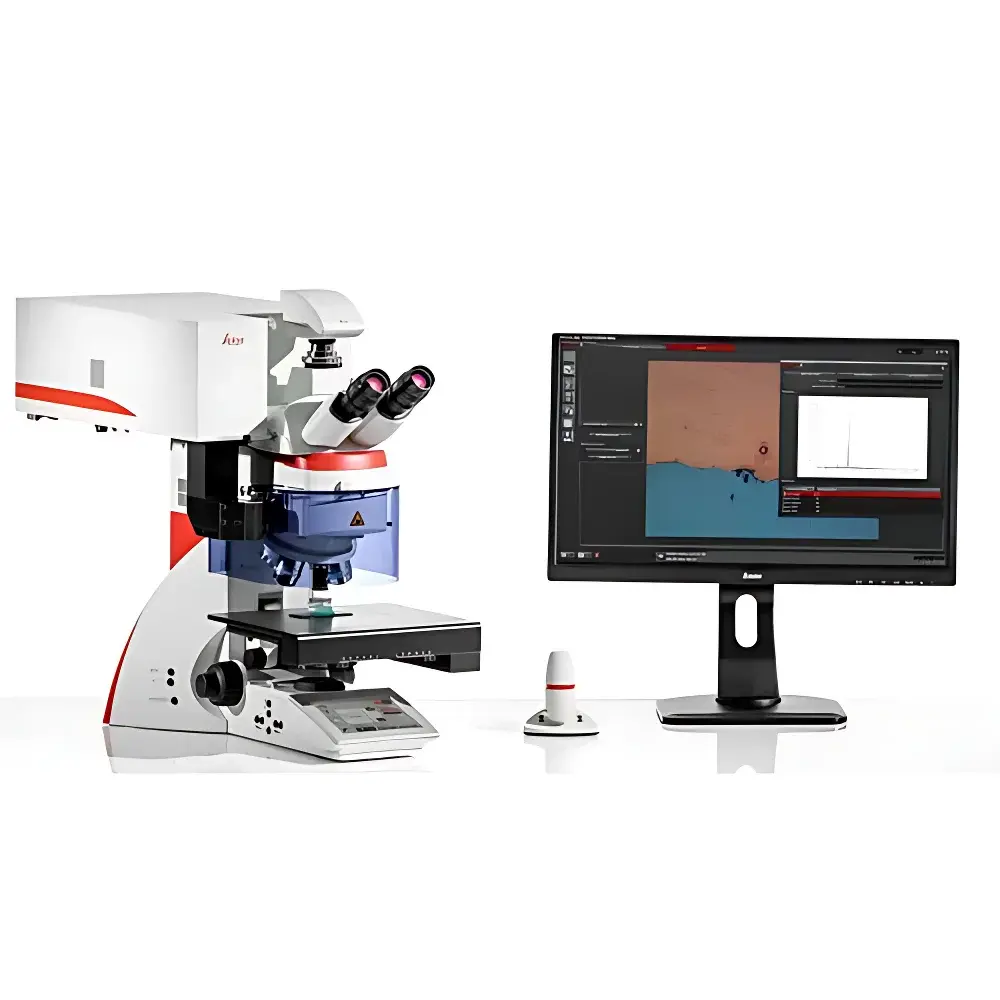
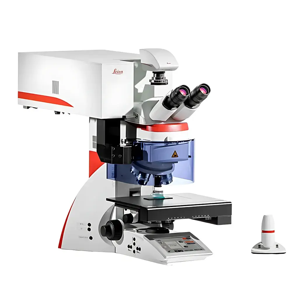
Leica DM6 M LIBS Materials Analysis Microscope
| Brand | Leica |
|---|---|
| Origin | Germany |
| Manufacturer Type | Authorized Distributor |
| Product Origin | Imported |
| Model | DM6 M LIBS Materials Analysis Microscope |
| Configuration | Upright |
| Integrated Imaging Analysis System | Yes |
| Total Magnification Range | 1.25× to 100× |
| Laser Induced Breakdown Spectroscopy (LIBS) Module | Integrated |
| Optical Contrast Methods | Brightfield, Darkfield, Polarization, Differential Interference Contrast (DIC), Interferometric Contrast (IC) |
| Sample Preparation | None Required |
| Analysis Speed | <1 second per spot |
| Depth Profiling Capability | Yes |
| Layer-by-Layer Elemental Mapping | Yes |
Overview
The Leica DM6 M LIBS Materials Analysis Microscope is an integrated upright metallurgical microscope engineered for simultaneous high-resolution structural imaging and rapid, in-situ elemental analysis via Laser-Induced Breakdown Spectroscopy (LIBS). Unlike conventional workflows requiring sample transfer between optical microscopy and electron-beam instruments (e.g., SEM-EDS), the DM6 M LIBS eliminates inter-system relocation, vacuum chamber loading, conductive coating, or beam alignment—enabling direct correlation of microstructural morphology and elemental composition within a single optical axis and coordinate system. Its core architecture leverages Leica’s precision mechanical stage, motorized objective turret, and multi-contrast illumination platform to deliver color-accurate, high-fidelity visualization across magnifications from 1.25× to 100×. The integrated LIBS module employs a pulsed Nd:YAG laser (typically 1064 nm, ns pulse duration) focused to a ~10–50 µm spot, generating transient microplasma on the sample surface; emitted atomic/ionic line spectra are collected via high-throughput Czerny–Turner spectrometer (200–800 nm range) and processed in real time using chemometric algorithms against reference spectral libraries.
Key Features
- Single-platform workflow: Co-registered optical imaging and LIBS spectroscopy without sample repositioning or relocation.
- No sample preparation required: Analyzes conductive and non-conductive solids—including metals, alloys, ceramics, composites, and coated substrates—in ambient air.
- Sub-second elemental identification: Qualitative and semi-quantitative detection of major, minor, and trace elements (Z ≥ 5, e.g., B, C, N, O, Na–U) per measurement.
- Depth profiling & layer-resolved analysis: Sequential laser ablation enables stratigraphic elemental mapping (e.g., paint multilayers, oxide films, diffusion zones).
- Multi-contrast optical imaging: Supports brightfield, darkfield, polarization, DIC, and Leica Interferometric Contrast (IC) for enhanced phase boundary and grain structure visualization.
- Motorized hardware coordination: Synchronized stage movement, focus, objective selection, and LIBS targeting ensure repeatable ROI registration across modalities.
- Modular upgrade path: Compatible with existing Leica DM6000 M or DM6 M metallurgical microscope platforms via certified field retrofit kit.
Sample Compatibility & Compliance
The DM6 M LIBS accommodates bulk solid specimens up to 150 mm × 100 mm × 50 mm (W × D × H) and thicknesses ≥ 0.1 mm. It supports flat, polished, rough, or as-received surfaces—including uncoated metallics, sintered powders, geological thin sections, and filtration media. No vacuum, cryo-cooling, or carbon/metal sputter coating is necessary. The system complies with IEC 61000-6-3 (EMC emission standards) and IEC 60825-1 (laser safety Class 4, fully interlocked enclosure). Data acquisition and reporting support audit trails aligned with GLP and GMP documentation requirements; optional software modules provide 21 CFR Part 11-compliant electronic signatures and user-access controls.
Software & Data Management
Leica Application Suite X (LAS X) serves as the unified control and analysis environment. It provides synchronized image capture, LIBS spectral acquisition, elemental distribution mapping (pixel-wise intensity integration), and depth-profile visualization. Spectral libraries include NIST Atomic Spectra Database (ASD) references, customizable user-defined material profiles, and auto-calibration routines using internal argon plasma lines. All raw spectra, metadata (laser energy, spot position, integration time), and processed reports are stored in vendor-neutral HDF5 format. Export options include CSV, TIFF (for overlay maps), and PDF reports compliant with ISO/IEC 17025 technical record requirements.
Applications
- Contamination source identification on filter membranes or component surfaces (e.g., automotive fuel filters, semiconductor wafer carriers).
- Rapid phase identification in multiphase alloys (e.g., austenite/ferrite ratio in stainless steels, intermetallics in Ni-superalloys).
- Coating integrity assessment: Thickness estimation, interdiffusion analysis, and delamination detection in PVD/CVD layers.
- Mineralogical characterization of ores and slags without polishing or embedding—preserving natural grain boundaries.
- Failure analysis: Corrosion product chemistry, inclusion composition mapping, and heat-affected zone (HAZ) elemental segregation.
- Quality control in additive manufacturing: Powder feedstock verification, melt pool residue analysis, and porosity-associated elemental segregation.
FAQ
Does the DM6 M LIBS require vacuum or special environmental conditions?
No. All analyses are performed in ambient laboratory air at standard temperature and pressure.
Can LIBS data be correlated quantitatively with certified reference materials?
Yes—semi-quantitative calibration is achievable using matrix-matched standards; full quantification requires empirical calibration curves per material class.
Is the laser safe for heat-sensitive or polymer-based samples?
The ns-pulsed ablation minimizes thermal diffusion; crater diameters remain sub-50 µm with negligible HAZ in most engineering polymers and composites.
How is spatial registration maintained between optical image and LIBS spot?
Via Leica’s motorized stage with 0.1 µm repeatability and hardware-triggered synchronization between camera exposure and laser firing.
Can the system export data for third-party chemometric software (e.g., MATLAB, Python scikit-learn)?
Yes—raw spectral ASCII/HDF5 exports enable custom PCA, PLS, or machine learning model development.