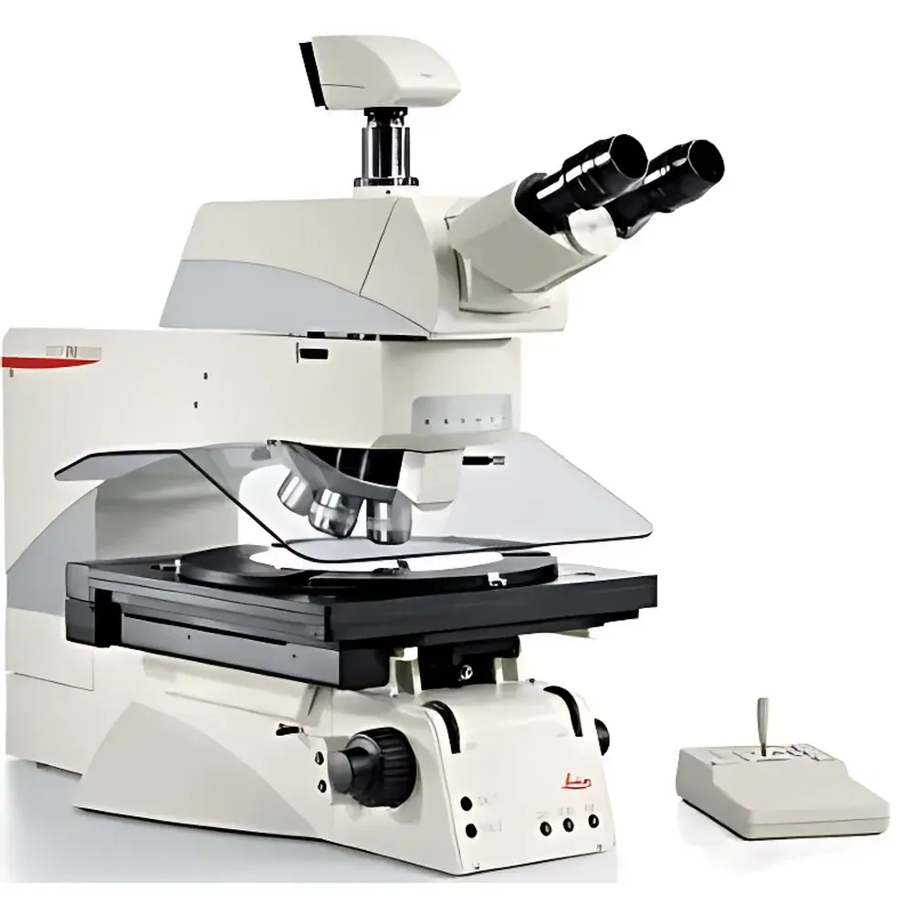
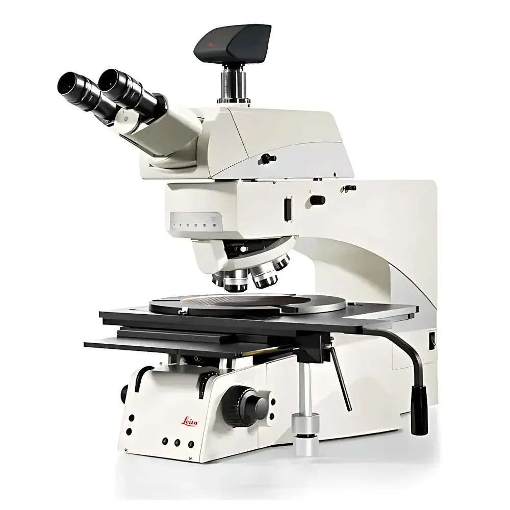
Leica DM12000M Advanced Intelligent Upright Materials Microscope
| Brand | Leica |
|---|---|
| Origin | Germany |
| Model | DM12000M |
| Configuration | Upright |
| Eyepiece Field Number | 25 mm |
| Objective Type | 32 mm Diameter Long Working Distance Industrial Objectives |
| Illumination | Integrated LED Reflected & Transmitted Light Source with Smart Intensity Control |
| Observation Modes | Brightfield, Darkfield, Polarization, Interference Contrast (DIC), Oblique Illumination |
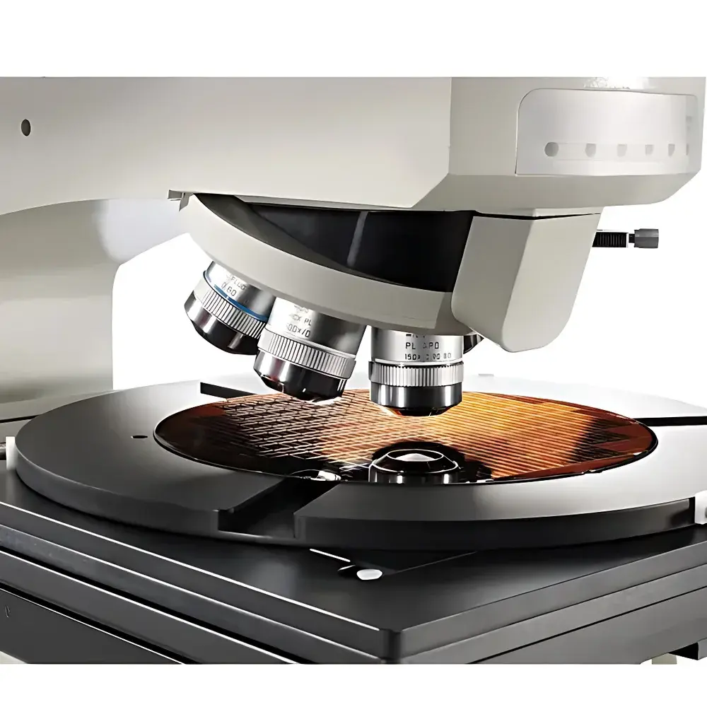
| Turret | Motorized 6-Position Nosepiece |
| Macro Objective | 0.7× Built-in Macro Lens for Wafer-Level Inspection |
| Stage | 12×12 inch Large Motorized/Manual Sample Stage |
| Optical Path | Apochromatic-Corrected, 25 mm Field of View Compatible |
| UV Capability | Optional High-Power UV LED Illumination (UV & OUV Modes) |
| Software Integration | Full Digital Imaging, Annotation, Calibration, and Metrology Support |
| Compliance | Designed for ISO 9001-aligned manufacturing environments |
Overview
The Leica DM12000M is an advanced intelligent upright materials microscope engineered for high-precision microstructural analysis across demanding industrial and research applications. Utilizing a fully apochromatic-corrected optical path optimized for a 25 mm field of view, the system delivers exceptional image fidelity, chromatic and spherical aberration correction, and consistent illumination uniformity—critical for quantitative metallurgical evaluation, semiconductor wafer inspection, and advanced materials characterization. Its upright configuration supports both reflected-light (epi-illumination) and transmitted-light observation modes, making it uniquely suited for opaque specimens such as metals, ceramics, and printed circuit boards, as well as semi-transparent samples including thin-film coatings and polymer laminates. The instrument’s core architecture integrates motorized optical components, intelligent illumination management, and modular expansion capabilities to meet evolving QA/QC, failure analysis, and R&D requirements in regulated environments.
Key Features
- Motorized 6-position objective nosepiece accommodating 32 mm diameter long working distance industrial objectives—enabling stable, vibration-resistant imaging of tall or topographically complex samples.
- Built-in 0.7× macro objective for rapid wafer-level survey imaging without stage repositioning—reducing inspection time and minimizing handling risk for fragile substrates.
- Smart LED illumination system with independent control of reflected and transmitted light intensity, aperture diaphragm, and condenser height—automatically recalling optimal settings per objective magnification and contrast mode.
- Full suite of contrast methods: brightfield, darkfield, polarization, differential interference contrast (DIC), and oblique illumination—supporting phase-sensitive defect detection, grain boundary delineation, and stress-induced birefringence analysis.
- 12 × 12 inch (305 × 305 mm) precision mechanical or motorized stage with fine positioning capability—designed for large-format specimens including 300 mm wafers, LCD panels, and composite cross-sections.
- Integrated color temperature stabilization system ensuring consistent white balance across illumination modes and exposure durations—essential for comparative image analysis and long-term measurement repeatability.
Sample Compatibility & Compliance
The DM12000M accommodates a broad range of solid-state materials—including ferrous and non-ferrous alloys, sintered ceramics, additive-manufactured metal parts, photomasks, solder joints, and particulate contaminants on cleanroom surfaces. Its long working distance optics and protective safety interlocks minimize contact risk during wafer or delicate component inspection. The platform conforms to ISO 10934-1 (microscopy terminology), ISO 9276-5 (particle size analysis by image analysis), and ASTM E3 (preparation and etching of metallographic specimens). When paired with Leica Application Suite X (LAS X) software configured for audit trail and electronic signature, the system supports compliance with FDA 21 CFR Part 11, EU Annex 11, and GLP/GMP documentation standards.
Software & Data Management
Leica LAS X software provides comprehensive digital microscopy functionality—including multi-channel image acquisition, real-time measurement (length, area, angle, particle count), automated focus mapping, and report generation with embedded calibration metadata. All hardware parameters (objective ID, illumination mode, magnification, aperture setting) are automatically embedded into TIFF or JPEG image headers. Optional modules enable automated multi-field-of-view stitching, inclusion of scale bars synchronized to objective magnification, and integration with LIMS or MES platforms via standardized API protocols. Raw image data remains unaltered; processing steps are non-destructive and logged with timestamped operator identification.
Applications
- Metallurgical quality assurance: grain size measurement (ASTM E112), inclusion rating (ASTM E45), porosity quantification, and heat-affected zone (HAZ) analysis.
- Semiconductor process control: die surface defect review, bond pad integrity verification, and lithography residue assessment under UV-enhanced contrast.
- Electronics reliability testing: solder joint void analysis, PCB trace continuity, and conformal coating uniformity evaluation.
- Advanced materials R&D: ceramic microcrack propagation studies, polymer phase separation imaging, and composite fiber orientation mapping.
- Failure analysis laboratories: root cause determination via comparative microstructural examination before/after environmental stress testing.
FAQ
Does the DM12000M support automated particle analysis for cleanliness testing?
Yes—when equipped with a motorized stage and LAS X Particle Analysis module, the system performs standardized ISO 16232 or VDA 19-compliant particle counting, sizing, and classification across multiple fields.
Can the UV illumination be used for fluorescence imaging?
No—the optional UV/OUV LED module is optimized for high-resolution reflectance imaging of sub-micron features (e.g., photoresist residues or oxide thickness variations); dedicated fluorescence modules require separate excitation filter sets and barrier filters.
Is remote operation supported?
Yes—via Ethernet-connected LAS X client-server deployment, enabling secure off-site microscope control, live streaming, and collaborative annotation from authorized workstations.
What documentation is provided for IQ/OQ validation?
Leica provides a comprehensive Validation Support Package including test protocols, acceptance criteria, and traceable calibration certificates for optical performance, stage positioning accuracy, and illumination stability—aligned with ISO/IEC 17025 laboratory accreditation requirements.