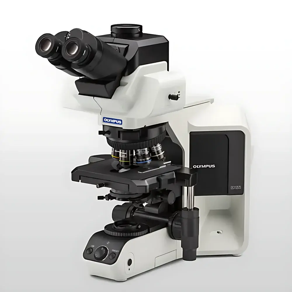
Olympus/EVIDENT BX53 LED Semi-Motorized Fluorescence Microscope
| Brand | Olympus/EVIDENT |
|---|---|
| Origin | Japan |
| Model | BX53 (LED) |
| Illumination Type | High-Stability White LED (Spectrally Optimized for Clinical Staining Contrast) |
| Optical Design | UIS2 Infinity-Corrected Optics |
| Fluorescence Capability | Standard Filter Cube Turret (6-position), Optional Motorized Turret |
| Focus Mechanism | Semi-motorized Z-axis (Manual Coarse/Fine + Motorized Precision Positioning) |
| Stage | Mechanical XY Stage with Vernier Scale |
| Compliance | CE, ISO 9001, JIS B 7070 |
Overview
The Olympus/EVIDENT BX53 LED Semi-Motorized Fluorescence Microscope is an engineered platform designed for routine and advanced light microscopy in clinical pathology, cytology, and academic life science laboratories. Built upon the proven UIS2 infinity-corrected optical system, the BX53 delivers high-fidelity color fidelity and consistent contrast across widefield brightfield, phase contrast, and fluorescence modalities. Its core illumination architecture integrates a spectrally optimized white LED light source—engineered to replicate the visible spectral distribution of traditional 12 V/100 W halogen lamps (peaking at ~3200 K correlated color temperature) while eliminating infrared heat emission and intensity drift. This spectral alignment ensures faithful rendering of critical diagnostic hues—including violet (e.g., DAPI-stained nuclei), cyan (e.g., FITC-labeled antibodies), and magenta (e.g., TRITC or Cy3 conjugates)—without requiring revalidation of existing staining protocols or subjective brightness recalibration. The semi-motorized configuration provides precise Z-axis repeatability (±0.1 µm step resolution) for multiplane imaging, z-stack acquisition, and longitudinal sample monitoring—while retaining manual coarse/fine focus ergonomics for rapid initial localization.
Key Features
- Spectrally matched white LED illumination (380–700 nm output) with <1% intensity fluctuation over 10,000 hours—eliminating lamp replacement cycles and thermal drift during extended imaging sessions.
- UIS2 optical system with apochromat-corrected objectives (10×–100× oil), delivering diffraction-limited resolution and minimal chromatic aberration across visible and near-UV excitation bands.
- Six-position filter cube turret (standard); optional motorized version enables automated multi-channel fluorescence switching under software control with <150 ms cube transition time.
- Semi-motorized Z-drive with programmable memory positions, enabling repeatable focal plane recall for time-lapse series, tissue section mapping, and QC slide re-examination.
- Ergonomic modular design: interchangeable observation tubes (trinocular, ergonomic inclined), integrated camera port (C-mount & F-mount options), and DIC/phase contrast compatibility without optical realignment.
- Compliance-ready architecture: supports audit trail logging (via optional cellSens software), user access levels, and electronic signature capability aligned with GLP/GMP documentation requirements.
Sample Compatibility & Compliance
The BX53 accommodates standard 1–3 mm thick glass microscope slides and coverslips (0.13–0.17 mm), including stained histopathology sections (H&E, PAS, Giemsa), immunofluorescent preparations, live-cell specimens in chambered coverslips, and whole-mount tissue slices up to 200 µm thickness. Its LED-based Köhler illumination maintains uniformity across 22 mm field number optics—critical for digital pathology scanning and quantitative morphometric analysis. The system conforms to IEC 61000-4 electromagnetic compatibility standards and carries CE marking per Directive 2014/30/EU (EMC) and 2014/65/EU (Medical Devices). All optical components are manufactured to JIS B 7070 specifications for microscope performance verification, and the platform supports validation documentation packages compliant with ISO/IEC 17025 and CLIA laboratory accreditation frameworks.
Software & Data Management
When paired with Olympus cellSens Dimension software (v3.1+), the BX53 enables full instrument control, multi-channel fluorescence acquisition, z-stack reconstruction, and quantitative intensity profiling (e.g., nuclear/cytoplasmic fluorescence ratio analysis). The software includes FDA 21 CFR Part 11-compliant modules: electronic signatures, audit trail generation (timestamped operator actions, parameter changes, image annotations), and role-based access control (administrator, technician, reviewer). Image metadata embeds objective magnification, exposure time, LED intensity setting, filter cube ID, and stage coordinates—enabling traceability from raw acquisition to final report. Export formats include TIFF (16-bit), OME-TIFF (for Bio-Formats compatibility), and PDF reports with embedded calibration certificates.
Applications
- Clinical cytology screening: high-throughput evaluation of Pap smears and fine-needle aspirates with enhanced contrast for low-contrast keratinized cells and metaplastic epithelium.
- Immunohistochemistry (IHC) assessment: reproducible quantification of membrane-bound (e.g., HER2) and nuclear (e.g., Ki-67) biomarker expression using standardized LED excitation and linear CCD response.
- Live-cell fluorescence monitoring: long-term timelapse of GFP/RFP-tagged proteins in adherent cultures—enabled by stable LED output and minimal phototoxicity versus mercury arc sources.
- Academic research: multiplexed fluorescence co-localization studies (e.g., synaptic protein clustering), mitotic index scoring, and morphological phenotyping in genetically modified model organisms.
- Quality assurance in biobanking: standardized imaging of frozen section controls across multiple instruments via calibrated LED intensity presets and objective-specific exposure profiles.
FAQ
Does the BX53 LED support UV excitation for DAPI staining?
Yes—the BX53’s LED illumination system emits down to 380 nm, and when used with a standard DAPI filter cube (excitation 350/50 nm, dichroic 395 nm, emission 460/50 nm), it delivers sufficient photon flux for robust nuclear counterstaining in fixed and permeabilized samples.
Can motorized functions be added post-purchase?
Yes—motorized Z-drive, filter turret, and XY stage modules are field-upgradable via certified service technicians; firmware and software licenses must be purchased separately.
Is the BX53 compatible with third-party cameras?
Yes—C-mount and F-mount mechanical interfaces support industry-standard scientific CMOS and sCMOS cameras; Olympus provides SDK documentation for custom API integration.
What maintenance is required for the LED illumination system?
No periodic lamp replacement or alignment is needed; annual intensity calibration using an NIST-traceable photodiode sensor is recommended for quantitative applications.
How does the BX53 meet regulatory requirements for clinical labs?
It supports full audit trail logging, user authentication, and electronic signatures via cellSens software—enabling compliance with CAP checklist MIC.40500, CLIA ’88, and ISO 15189:2022 clause 5.9.2 for result verification and data integrity.


