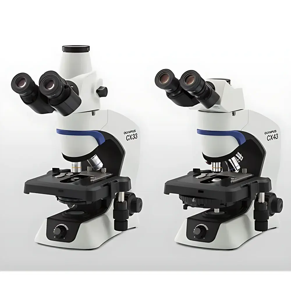
Olympus EVIDENT CX43 and CX33 Biological Microscopes
| Brand | Olympus/EVIDENT |
|---|---|
| Origin | Japan |
| Model | CX43 / CX33 |
| Import Status | Imported Instrument |
| Distribution Type | Authorized Distributor (Non-Manufacturer) |
| Pricing | Available Upon Request |
Overview
The Olympus EVIDENT CX43 and CX33 are upright, routine biological microscopes engineered for high-throughput educational laboratories, clinical pathology units, and quality control environments in life science research. Designed around a modular, ergonomic optical architecture, both models utilize infinity-corrected optical systems with high-transmission, anti-reflective coated optics to deliver consistent contrast, color fidelity, and resolution across wide-field viewing. The CX43 features an advanced UIS2 optical platform with enhanced Köhler illumination uniformity and optional phase contrast capabilities, while the CX33 provides a streamlined, cost-optimized configuration with fixed magnification objectives and integrated LED illumination—ensuring stable, flicker-free brightness over extended operational cycles. Neither system incorporates fluorescence or confocal imaging modules; they are purpose-built for brightfield, phase contrast, and basic polarized light observation of fixed or live unstained specimens on standard glass slides.
Key Features
- Ergonomic design with adjustable interpupillary distance (55–75 mm), inclined 30° binocular tube, and low-positioned focusing controls to reduce operator fatigue during prolonged use.
- Infinity-corrected optical path (180 mm tube length) compatible with standardized UIS2 objective lenses (4×, 10×, 40×, 100× oil immersion), supporting parfocal and parcentric alignment.
- Dual-LED illumination system (CX43) or single high-efficiency white LED (CX33) with intensity control dial and built-in field diaphragm—providing >10,000 hours of stable output and compliance with IEC 62471 photobiological safety standards.
- Reinforced metal frame construction with vibration-damped base and coaxial coarse/fine focusing mechanism (2 mm coarse travel, 0.002 mm fine increment) for precise Z-axis positioning.
- CX43-specific enhancements: Adjustable condenser (NA 1.25) with centering screws, optional sliding swing-out phase telescope, and interchangeable eyepiece tubes supporting 10×/15× wide-field oculars and camera port integration.
Sample Compatibility & Compliance
These microscopes accommodate standard 1″ × 3″ (25 × 75 mm) microscope slides and cover glasses up to 0.17 mm thickness. Specimen height clearance allows for routine histology sections, blood smears, microbiological cultures on agar, and live-cell observation in shallow Petri dishes (≤15 mm depth). Both models comply with ISO 10934-1:2002 (Microscopes — Nomenclature of components) and meet CE marking requirements under the EU Medical Device Regulation (MDR 2017/745) as Class I non-invasive diagnostic equipment. They support GLP-compliant documentation when paired with certified digital imaging systems (e.g., Olympus UC50 camera + cellSens software), though standalone operation does not require FDA 21 CFR Part 11 validation.
Software & Data Management
The CX43 and CX33 operate independently of proprietary software but are fully compatible with Olympus’ cellSens imaging suite (v2.4 or later) via USB 3.0 or HDMI output when connected to a compliant C-mount adapter and digital camera module (e.g., DP28, UC50). cellSens supports annotation, multi-channel overlay, measurement calibration (µm/pixel), time-lapse capture, and export in TIFF, JPEG2000, and OME-TIFF formats—enabling traceable image archiving aligned with institutional data retention policies. Raw image metadata (objective magnification, exposure time, illumination intensity) is embedded per DICOM Supplement 146 conventions where applicable. No onboard storage or network connectivity is integrated; all data handling occurs externally through validated laboratory IT infrastructure.
Applications
- Undergraduate and graduate-level biology instruction: mitosis staging, plant tissue anatomy, protozoan motility analysis.
- Clinical hematology: peripheral blood smear differential counts, reticulocyte assessment, and malaria parasite identification (using Giemsa-stained thin films).
- Pharmaceutical QC: particulate inspection in parenteral solutions, filter membrane analysis, and excipient morphology verification.
- Food microbiology: detection of Salmonella, Listeria, and yeast contamination in enrichment broths using Gram staining protocols.
- Agricultural diagnostics: fungal spore morphology, nematode identification, and pollen grain characterization in botanical reference labs.
FAQ
Are the CX43 and CX33 suitable for fluorescence microscopy?
No. These models lack mercury/xenon lamp housings, excitation/emission filter cubes, and dichroic mirrors required for epifluorescence. Fluorescence capability is available only on Olympus BX series or CKX inverted platforms.
Can I upgrade a CX33 to CX43 functionality?
No. Core optical and mechanical components—including condenser design, focusing mechanism, and tube lens configuration—are not cross-compatible between the two models. Upgrades are limited to accessories such as eyepieces, objectives, or LED intensity controllers.
What is the warranty coverage for imported CX-series microscopes?
Olympus EVIDENT provides a standard 2-year limited warranty covering parts and labor for defects in materials and workmanship, valid globally under authorized distributor service networks. Extended service plans (up to 5 years) are available upon registration.
Do these microscopes meet ISO/IEC 17025 requirements for accredited testing labs?
Yes—when operated with calibrated stage micrometers and traceable reference standards, and when maintenance logs, user training records, and performance verification reports (e.g., resolution test using USAF 1951 target) are maintained per Clause 6.4 and Annex A.3 of ISO/IEC 17025:2017.
Related Products


