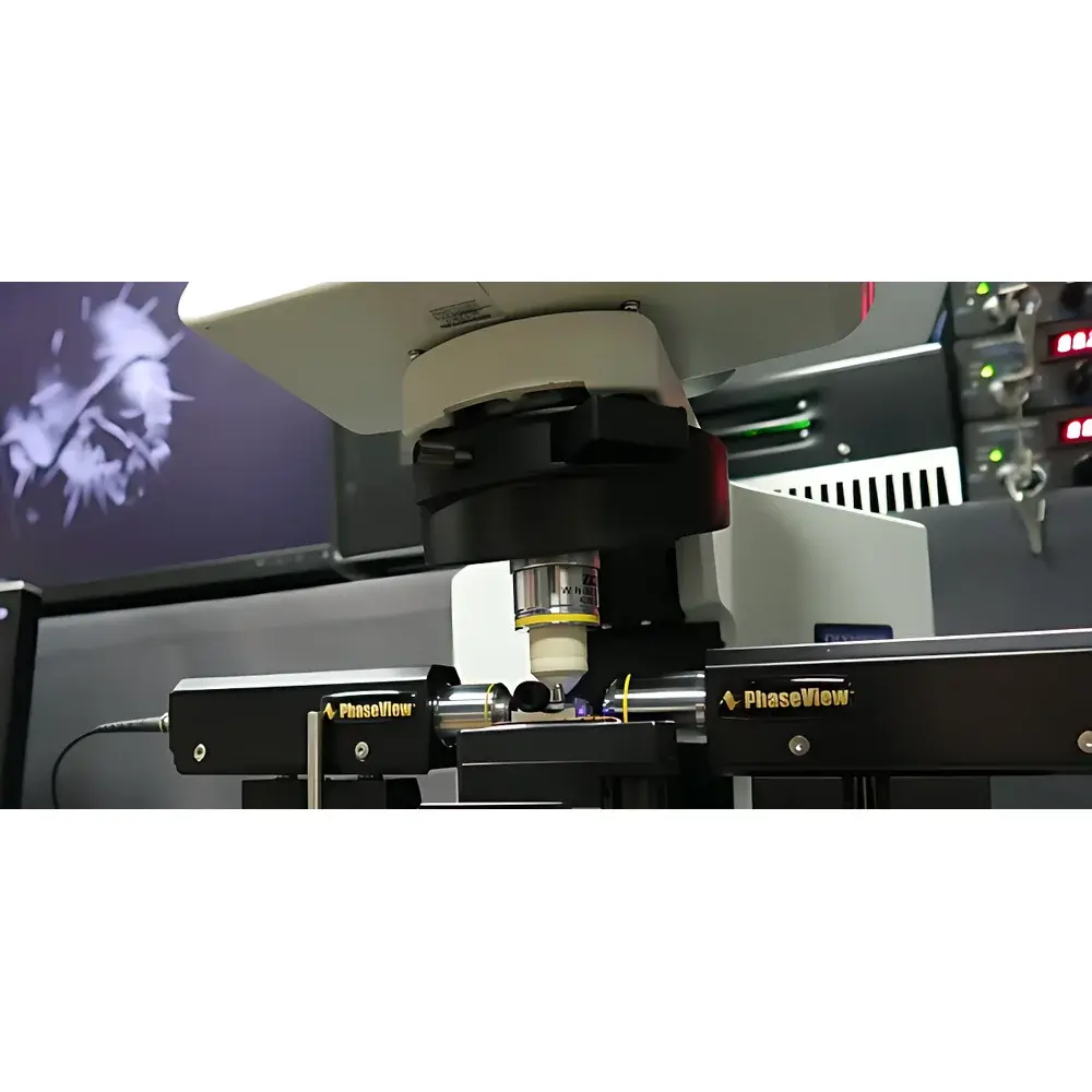
PhaseView Alpha Laser Light-Sheet Microscopy System
| Brand | PhaseView |
|---|---|
| Origin | France |
| Model | Alpha |
| Scanning Module | High-Speed Laser Light-Sheet Scanner |
| Scanning Method | Remote Digital Focus |
| Optical Zoom | 10× |
| Excitation Wavelength Range | 375–785 nm |
| Objective | Standard 10× 0.25 NA Air Objective (Optional: 5×, 20×) |
| Detection Optics | Finite–Infinite Type, Air/Water-Dipping Lenses |
| Volumetric Scanning Modes | Motorized Sample Scan (25 fps), NeoScan Remote Focus (25 fps), ThunderScan Ultra-High-Speed Remote Focus (100 fps) |
| Compatible Detectors | Scientific sCMOS Cameras (Large-Format) |
| Sample Volume Capacity | Up to 10 mm × 10 mm × 10 mm |
| Illumination Geometry | Orthogonal Light-Sheet Excitation / Perpendicular Emission Detection |
Overview
The PhaseView Alpha Laser Light-Sheet Microscopy System is a high-performance, turnkey light-sheet fluorescence microscope engineered for quantitative 3D imaging of live, large-volume biological specimens with minimal phototoxicity and exceptional spatiotemporal fidelity. Built upon PhaseView’s proprietary remote digital focusing technology and integrated laser light-sheet illumination architecture, the Alpha system enables optical sectioning via a thin, planar sheet of excitation light—orthogonally oriented relative to the detection axis—thereby decoupling illumination from signal collection. This orthogonal geometry eliminates out-of-focus excitation and significantly reduces photobleaching and photodamage compared to point-scanning modalities such as confocal or multiphoton microscopy. The system operates across a broad excitation spectrum (375–785 nm), supporting multicolor labeling with standard fluorophores (e.g., DAPI, GFP, mCherry, Cy5) and near-infrared probes. Designed for reproducible, long-term time-lapse experiments, the Alpha delivers subcellular resolution within centimeter-scale samples—making it especially suited for developmental biology, neuroimaging, immunology, and organ-level functional studies in intact model organisms.
Key Features
- Patented remote digital focus optics enabling diffraction-limited axial scanning without mechanical objective movement—eliminating vibration-induced image drift and preserving spatial registration across Z-stacks.
- High-speed volumetric acquisition: ThunderScan mode achieves up to 100 optical sections per second, facilitating real-time tracking of dynamic processes including cardiomyocyte contraction, neural calcium transients, and vascular perfusion.
- Modular laser excitation platform supporting continuous-wave (CW) fiber lasers, laser diodes, or DPSS sources; wavelength selection from deep UV (375 nm) to near-IR (785 nm) ensures compatibility with diverse fluorophore families and optogenetic actuators.
- Dual-arm illumination configuration (standard single-arm, optional dual-arm) for symmetric light-sheet generation, improving uniformity and reducing shadow artifacts in opaque or scattering specimens.
- Optimized optical train featuring finite–infinite correction lenses and interchangeable air- or water-dipping objectives (standard 10× 0.25 NA; optional 5× and 20×), enabling flexible magnification and immersion matching for varied sample refractive indices.
- Native integration with scientific-grade sCMOS cameras—including large-format sensors—ensuring high quantum efficiency, low read noise, and wide field-of-view coverage for comprehensive volumetric sampling.
Sample Compatibility & Compliance
The PhaseView Alpha accommodates native, cleared, or lightly embedded specimens up to 10 mm × 10 mm × 10 mm in volume—including whole zebrafish embryos, Drosophila larvae, C. elegans, mouse brain slices, explanted lymph nodes, embryonic hearts, ocular tissues, and 3D organoid cultures. Its non-invasive illumination strategy complies with best practices for live-sample integrity under GLP-aligned experimental protocols. The system supports common tissue-clearing methods (e.g., CLARITY, iDISCO, BABB) and is compatible with standard mounting media (e.g., FocusClear, RIMS). Data acquisition metadata—including timestamp, laser power, Z-step size, exposure duration, and objective ID—is automatically embedded in TIFF/OME-TIFF files, ensuring traceability for regulatory-compliant workflows aligned with ISO/IEC 17025 documentation standards and FDA 21 CFR Part 11 audit-trail requirements when paired with validated software environments.
Software & Data Management
The Alpha system ships with PhaseView’s unified acquisition and reconstruction suite, supporting synchronized control of illumination, remote focus, stage motion, and camera triggering. Core capabilities include real-time preview, multi-channel time-lapse capture, automated Z-stack acquisition, and hardware-accelerated deconvolution using measured PSF models. Post-acquisition tools enable volumetric rendering (maximum intensity projection, volume rendering, surface reconstruction), quantitative morphometry (region-based segmentation, distance mapping, colocalization analysis), and 4D visualization (3D + time). Export formats include OME-TIFF, HDF5, and N5 for interoperability with open-source platforms (e.g., Fiji/ImageJ, Napari, BigDataViewer) and institutional HPC pipelines. Optional modules support AI-assisted segmentation (via trained U-Net models) and batch processing compliant with FAIR data principles.
Applications
- Developmental biology: Long-term, high-resolution imaging of embryogenesis in zebrafish and Drosophila, capturing cell lineage dynamics, tissue folding, and morphogen gradient formation.
- Neuroscience: Whole-brain functional imaging in larval zebrafish and mouse cortical slabs using GCaMP or jRGECO reporters; structural mapping of synaptic density and axonal arborization.
- Immunology: 3D visualization of antigen trafficking, T-cell motility within lymph nodes, and germinal center dynamics in intact tissue explants.
- Cardiovascular research: Quantitative assessment of cardiac wall motion, valve kinetics, and hemodynamic flow patterns in beating embryonic hearts.
- Marine and plant biology: Low-light imaging of fragile planktonic organisms (e.g., ascidians, cephalopod embryos) and time-resolved monitoring of stomatal conductance or root gravitropism in light-sensitive seedlings.
- 3D cell culture: Phenotypic screening of tumor spheroids and iPSC-derived organoids under physiological oxygen and nutrient gradients.
FAQ
What distinguishes light-sheet microscopy from confocal or spinning-disk systems?
Light-sheet microscopy illuminates only the focal plane being imaged, drastically reducing total photon dose and enabling longer viability in live specimens—unlike point-scanning techniques that expose the entire specimen volume to excitation light.
Can the Alpha system be integrated into existing microscope frameworks?
Yes—the Alpha is available in both standalone and modular configurations; its optical paths and control APIs are designed for integration with motorized stages, environmental chambers, and third-party electrophysiology rigs.
Is ThunderScan compatible with adaptive optics correction?
ThunderScan’s remote focus mechanism is fully compatible with deformable mirror-based aberration correction when used with wavefront sensing modules and closed-loop calibration routines.
Does the system support two-photon light-sheet excitation?
While the standard Alpha configuration uses single-photon excitation, custom variants with pulsed IR lasers and harmonic generation optics can be configured upon request for deep-tissue two-photon light-sheet applications.
How is calibration performed for quantitative intensity measurements?
The system includes factory-calibrated photometric references and supports user-performed flat-field correction, laser power monitoring via inline photodiodes, and emission spectral unmixing using reference spectra libraries.


