3i Vivo SDC Upright In Vivo Spinning Disk Confocal Microscopy System
| Brand | 3i |
|---|---|
| Origin | USA |
| Manufacturer Type | Authorized Distributor |
| Origin Category | Imported |
| Model | Vivo SDC |
| Pricing | Upon Request |
Overview
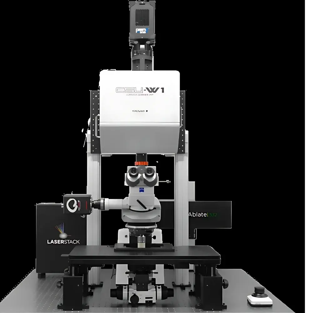
The 3i Vivo SDC Upright In Vivo Spinning Disk Confocal Microscopy System is an engineered platform for high-fidelity, long-term, three-dimensional intravital imaging of live small-animal models and intact tissues. Designed specifically for physiological relevance in dynamic biological contexts, the system integrates Vector2 dual-microlens spinning disk confocal technology with precision in vivo environmental control—addressing the persistent experimental constraints of sample viability maintenance, imaging depth limitation, and temporal resolution in live-tissue observation. Unlike inverted configurations optimized for cultured cells, the upright architecture enables direct compatibility with surgically prepared preparations—including cranial windows, mesenteric beds, dorsal skinfold chambers, and femoral artery exposures—without requiring tissue clearing, sectioning, or mounting media that compromise native physiology. The system operates on the principle of parallelized optical sectioning via high-speed rotating microlens arrays, delivering diffraction-limited resolution with minimal phototoxicity and photobleaching, making it suitable for millisecond-to-multi-hour acquisition protocols across neurovascular, oncological, immunological, and developmental research domains.
Key Features
- Vector2 Spinning Disk Confocal Engine: Dual-microlens architecture enabling simultaneous excitation and emission path optimization; supports both one-photon and two-photon imaging modalities for adaptive depth penetration up to 1 mm in scattering tissue, with optional adaptive optics integration.
- In Vivo Environmental Stabilization: Integrated surgical-stage climate control system regulating localized temperature (±0.1 °C), humidity, and gas composition (O2/CO2/N2 mixtures); coupled with hardware-based autofocus stabilization to compensate for respiratory and cardiac motion drift over extended time-lapse sessions.
- Multiwavelength Laser Ablation & Optogenetic Control: Onboard 355 nm and 532 nm pulsed lasers for subcellular ablation, endothelial injury induction, and thrombus initiation; fully synchronized with patterned illumination modules supporting computer-generated holography (CGH) for spatially resolved optogenetic activation, photoconversion, and photolysis.
- High-Speed Dynamic Acquisition: sCMOS camera interface with >95% quantum efficiency and sub-10 ms frame latency; combined with galvo-assisted laser line switching and real-time shutter synchronization for capturing rapid physiological events—e.g., platelet adhesion kinetics, leukocyte rolling velocity, capillary perfusion dynamics.
- Unified Hardware–Software Orchestration: SlideBook 6 software provides deterministic timing control across all subsystems—laser firing, stage movement, focus correction, camera exposure, and light patterning—enabling reproducible, timestamp-synchronized multimodal experiments compliant with GLP/GMP data integrity standards.
Sample Compatibility & Compliance
The Vivo SDC accommodates live murine and rat models under anesthesia-compatible physiological monitoring (ECG, respiration, core temperature). Its upright geometry and large working distance water-immersion objectives (e.g., 25×/1.10 NA, 40×/0.80 NA) allow unobstructed access to surgically exposed tissues while maintaining immersion medium stability. The system conforms to ISO 13485–aligned manufacturing practices and supports audit-ready data provenance through SlideBook’s built-in electronic lab notebook (ELN) features—including user authentication, version-controlled acquisition protocols, and FDA 21 CFR Part 11–compliant audit trails when deployed in regulated preclinical environments.
Software & Data Management
SlideBook 6 serves as the central analytical and control environment, offering modular workflows for deconvolution (Wiener and constrained iterative algorithms), volumetric rendering (ray-casting and maximum-intensity projection), spatiotemporal registration, and quantitative morphometric analysis (vessel diameter, branching density, fluorescence intensity kinetics). Raw data are stored in standardized OME-TIFF format with embedded metadata (acquisition parameters, laser power, objective ID, environmental logs), ensuring FAIR (Findable, Accessible, Interoperable, Reusable) compliance. Batch processing pipelines support GPU-accelerated denoising and motion correction, scalable to terabyte-scale intravital datasets generated during multi-day longitudinal studies.
Applications
- Vascular Biology & Thrombosis Research: Real-time quantification of thrombus nucleation, growth, and lysis within microvasculature; visualization of neutrophil extracellular trap (NET) deployment and endothelial glycocalyx shedding under inflammatory challenge.
- Neuroscience: Longitudinal tracking of dendritic spine turnover, calcium transient propagation in cortical layers, and axonal transport dynamics in awake or anesthetized rodents using chronic cranial windows.
- Intravital Oncology: Monitoring tumor angiogenesis, immune cell infiltration (T-cell motility, macrophage phagocytosis), and metastatic seeding in orthotopic and transgenic models; paired with photodynamic ablation to assess local immune response.
- Optogenetics & Cell-Specific Perturbation: Closed-loop experiments combining functional readout (Ca2+ or voltage indicators) with targeted perturbation (ChR2 activation, Caspase-3 photoactivation) at single-cell resolution in intact tissue.
- Pharmacokinetic/Pharmacodynamic Assessment: In vivo evaluation of drug biodistribution, target engagement, and functional impact on vascular permeability, neuronal excitability, or tumor metabolism—bridging in vitro screening and clinical translation.
FAQ
What animal models are supported?
The system is validated for mice, rats, zebrafish larvae, and Drosophila pupae; custom stage adapters are available for larger models.
Can the system perform two-photon imaging without hardware modification?
Yes—integrated dual-modality capability allows seamless switching between one-photon spinning disk and two-photon excitation using the same optical path and objective turret.
Is SlideBook software compatible with third-party analysis tools?
SlideBook exports OME-TIFF, HDF5, and N5 formats; Python and MATLAB APIs enable integration with scikit-image, napari, and custom deep-learning pipelines.
How is phototoxicity minimized during prolonged imaging?
The spinning disk architecture reduces peak irradiance by >100× compared to point-scanning confocal systems; combined with precise laser power modulation and adaptive dwell-time control, it enables >8-hour continuous imaging without measurable loss of cellular viability.
Does the system support regulatory-compliant data archiving?
Yes—SlideBook 6 includes configurable audit trail logging, digital signature authentication, and encrypted database storage aligned with 21 CFR Part 11 and ISO/IEC 17025 requirements for GLP-compliant laboratories.


