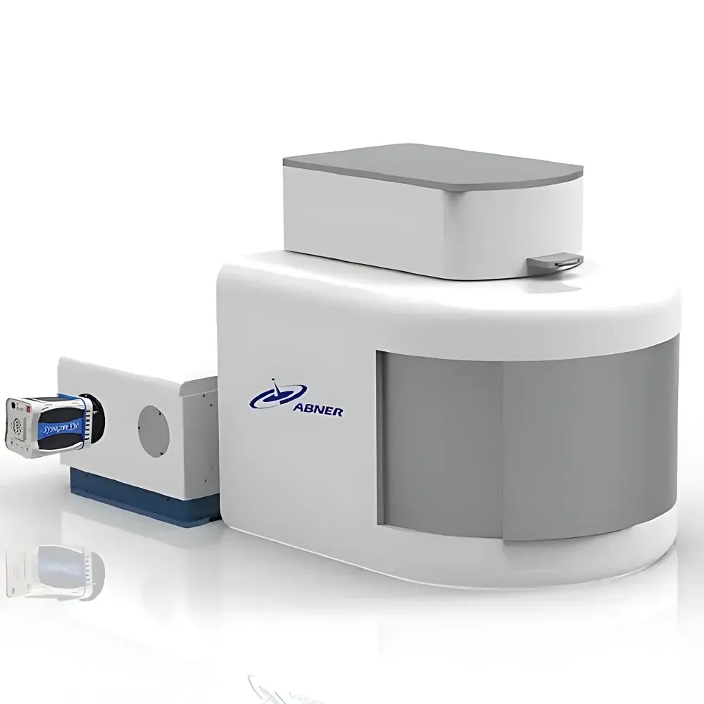
Abner ABN-CRM-001 Laser Confocal Raman Microscope
| Brand | Abner |
|---|---|
| Origin | Jiangsu, China |
| Model | ABN-CRM-001 |
| Instrument Type | Point-Scanning Confocal Microscope |
| Lasers | Standard integrated 445 nm, 532 nm, and 633 nm diode lasers |
| Detector | EMCCD sensor with 1600 × 200 or 1600 × 400 pixel array |
| Scanning Module | High-resolution Nikon CCD camera for real-time image display and digital storage |
| Focusing Mechanism | Precise manual adjustment of eyepiece and objective lens to locate micron-scale samples and visually confirm laser focus position on target particles |
Overview
The Abner ABN-CRM-001 Laser Confocal Raman Microscope is a fully integrated multimodal imaging platform engineered for correlative structural, functional, and molecular analysis of biological specimens at submicron spatial resolution. Combining confocal fluorescence microscopy, high-sensitivity Raman spectroscopy, and precision point-scanning architecture in a single optical train, the system enables simultaneous acquisition of morphological contrast (via reflected/transmitted light), functional localization (via multi-channel fluorescence), and label-free chemical identification (via vibrational Raman signatures). Its core optical design follows the principle of pinhole-confined detection—where only in-focus photons pass through a spatially aligned confocal aperture—thereby rejecting out-of-plane scattered light and delivering optical sectioning capability essential for 3D reconstruction of thick tissues, live cells, and heterogeneous biomaterials. The instrument operates under diffraction-limited conditions across visible excitation bands and is optimized for low-phototoxicity operation, making it suitable for extended time-lapse studies of viable specimens.
Key Features
- Confocal optical architecture with adjustable pinhole alignment for optimal axial resolution and signal-to-noise ratio in both fluorescence and Raman modalities
- Triple-wavelength laser excitation (445 nm, 532 nm, 633 nm) supporting broad-spectrum fluorophore compatibility (e.g., DAPI, FITC, TRITC, Cy5) and resonance-enhanced Raman acquisition
- Back-illuminated EMCCD detector with electron-multiplying gain control, enabling single-photon-level sensitivity for weak Raman signals from native biomolecules
- Point-scanning Raman mapping capability with user-defined XY grid parameters (start/stop coordinates, step size), synchronized with real-time positional feedback
- Unified coordinate system across all imaging modes ensures pixel-perfect spatial registration between fluorescence images, confocal Z-stacks, and Raman hyperspectral cubes
- Modular optical path design supports future integration of additional lasers (e.g., 785 nm for reduced autofluorescence), spectral filters, or polarization optics
Sample Compatibility & Compliance
The ABN-CRM-001 accommodates standard glass-bottom petri dishes, microscope slides, tissue sections (frozen or FFPE), hydrogels, electrospun scaffolds, and cultured 2D/3D cell models. Sample mounting requires no conductive coating or vacuum environment, preserving native biochemical integrity. The system adheres to ISO 10993-5 biocompatibility guidelines for optical interface materials and meets CE marking requirements for laboratory instrumentation (2014/30/EU EMC Directive and 2014/35/EU LVD). All software operations comply with ALCOA+ principles for data integrity, supporting audit trails, electronic signatures, and role-based access control per FDA 21 CFR Part 11 when deployed in regulated environments (e.g., preclinical GLP studies).
Software & Data Management
The proprietary Abner RamanSuite v3.2 software provides end-to-end workflow management—from hardware synchronization and acquisition control to multivariate spectral analysis and 3D visualization. It incorporates chemometric algorithms including PCA, cluster analysis (k-means, hierarchical), and supervised classification (SVM, PLS-DA) trained on an internal spectral library covering >1,000 endogenous biomolecules (proteins, lipids, nucleic acids, metabolites). Quantitative mapping features enable concentration profiling based on band intensity calibration curves, with uncertainty estimation propagated from spectral noise statistics. Raw data are stored in vendor-neutral HDF5 format with embedded metadata (wavelength calibration, laser power, integration time, objective magnification), ensuring long-term reproducibility and third-party interoperability (e.g., with Python-based scikit-learn or MATLAB toolboxes).
Applications
- Cellular biochemistry: Spatially resolved mapping of protein conformational states, lipid phase segregation, and redox status in live neurons or immune cells without exogenous labels
- Tissue pathology: Discrimination of tumor margins in unstained surgical specimens via Raman spectral fingerprints correlated with H&E-aligned confocal morphology
- Drug delivery research: Co-localization of fluorescently tagged therapeutics with intracellular Raman markers of apoptosis or lysosomal activity
- Biomaterial characterization: In situ monitoring of collagen crosslinking, mineral deposition, or polymer degradation kinetics within 3D scaffolds under physiological conditions
- Microbiome analysis: Identification of bacterial species and metabolic phenotypes directly in biofilm matrices using strain-specific Raman bands
FAQ
What is the spatial resolution limit for Raman mapping on this system?
Under 532 nm excitation with a 100× oil-immersion objective (NA 1.4), the lateral resolution is approximately 250 nm (FWHM), consistent with the diffraction limit. Axial resolution in confocal Raman mode is ~600 nm.
Can the system perform time-resolved Raman measurements?
Yes—the software supports gated acquisition with microsecond temporal resolution using external TTL triggers, enabling pump-probe or kinetic Raman studies of fast biochemical transitions.
Is spectral calibration traceable to NIST standards?
All spectral calibrations are performed using certified polystyrene and silicon reference standards (NIST SRM 2241 and SRM 2242), with daily automated validation routines logged in the audit trail.
How is photodamage minimized during live-cell Raman imaging?
Laser power is dynamically controlled below 100 µW at the sample plane; exposure time per pixel is optimized using adaptive dwell-time algorithms that maintain SNR while limiting cumulative fluence to <1 J/cm².
Does the system support remote operation and data sharing?
Yes—RamanSuite includes secure HTTPS-based web interface for remote monitoring, real-time collaboration, and encrypted cloud backup of spectral datasets compliant with GDPR and HIPAA data handling protocols.


