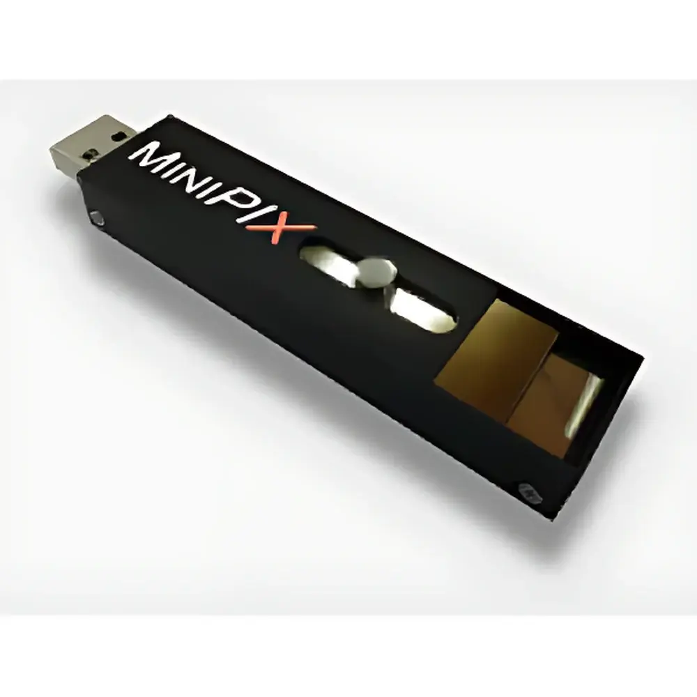
Advacam MiniPIX TPX Photon-Counting X-ray Detector
| Brand | Advacam |
|---|---|
| Origin | Czech Republic |
| Model | MiniPIX TPX |
| Detector Chip | Timepix (256 × 256 pixels) |
| Pixel Size | 55 µm |
| Sensor Material | Silicon |
| Sensor Thickness | 300 µm |
| Readout Interface | USB 2.0 |
| Frame Rate | up to 30 fps (1 ms exposure) |
| Operating Systems | Windows, macOS, Linux |
| Software | PIXET Lite (included), optional PIXET Pro or Pixelman |
| Radiation Types Detected | X-rays, neutrons, alpha particles, electrons, ions |
| Energy-resolved Imaging | Yes |
| Spatial Resolution | 55 µm |
| Dynamic Range | Effectively unlimited (zero dark current, photon-counting operation) |
Overview
The Advacam MiniPIX TPX is a compact, single-chip photon-counting X-ray detector engineered for high-fidelity radiation imaging and spectroscopic analysis. Built around the hybrid pixelated Timepix ASIC—bonded to a 300 µm thick silicon sensor—it operates on the principle of per-pixel time-over-threshold (ToT) measurement, enabling energy-sensitive detection and discrimination of individual incident quanta across multiple radiation modalities. Unlike integrating detectors, the MiniPIX TPX employs event-driven readout with zero dark current, delivering true digital counting performance without readout noise or gain drift. Its 256 × 256 pixel array (55 µm pitch) provides intrinsic spatial resolution ideal for micro-radiography, diffraction pattern analysis, and particle tracking. The device is designed for laboratory, educational, and field-deployable applications where portability, low-power operation (<2 W), and real-time spectral imaging are critical—such as in synchrotron beamlines, neutron facilities, nuclear safeguards, and materials stress mapping.
Key Features
- Photon-counting architecture: Each pixel independently registers and energy-tags individual photons or particles, eliminating integration artifacts and enabling quantitative fluence mapping.
- 55 µm spatial resolution: Defined by pixel pitch and charge sharing correction algorithms, supporting sub-millimeter feature resolution in high-magnification X-ray or neutron imaging setups.
- Energy-resolved imaging (spectral imaging): Time-over-threshold mode allows estimation of deposited energy per pixel—enabling material discrimination, K-edge imaging, and background rejection in mixed-field environments.
- Multi-radiation compatibility: Validated for X-rays (5–60 keV), thermal/cold neutrons (via 6LiF or 10B converters), alpha particles, electrons, and heavy ions—facilitating cross-platform use in radiation physics labs.
- Compact, embeddable form factor: Dimensions of 45 × 45 × 25 mm (excluding USB cable) permit integration into confined geometries—including pipe inspection rigs, portable neutron radiography carts, and custom collimator assemblies.
- USB 2.0 interface with deterministic timing: Sustains up to 30 full-frame acquisitions per second at 1 ms exposure, with hardware-triggered synchronization for pump-probe or time-resolved experiments.
Sample Compatibility & Compliance
The MiniPIX TPX is compatible with standard X-ray optics—including pinhole apertures, polycapillary lenses, and zone plates—as well as neutron converter foils mounted directly on the sensor housing. Its radiation hardness (tested up to 109 protons/cm2) and stable response over temperature (15–30 °C ambient) support long-duration monitoring in regulated environments. While not certified as a medical device, the system complies with EU Electromagnetic Compatibility Directive 2014/30/EU and RoHS 2011/65/EU. Data acquisition workflows using PIXET software support audit-ready metadata logging (timestamp, exposure, bias voltage, threshold settings), aligning with GLP documentation requirements for radiation safety and QA/QC protocols.
Software & Data Management
Control and analysis are performed via PIXET Lite (freely distributed, supports Windows/macOS/Linux), offering live histogramming, cluster analysis, energy calibration tools, and TIFF/RAW export. Optional PIXET Pro adds batch processing, tomographic reconstruction pipelines (FBP, SART), and scripting interfaces (Python API). Pixelman provides low-level register access for advanced users developing custom triggering or firmware-modified acquisition modes. All software versions enforce consistent file naming, embedded EXIF-style headers, and lossless 16-bit integer storage—ensuring traceability for ISO/IEC 17025-accredited laboratories. Exported spectra and image stacks are compatible with third-party platforms including MATLAB, ImageJ/Fiji, and ROOT for statistical modeling and Monte Carlo validation.
Applications
- X-ray diffraction (XRD) pattern acquisition from micro-crystalline samples, especially in transmission geometry with energy discrimination to suppress fluorescence background.
- Residual stress mapping in engineering alloys using energy-resolved Laue or monochromatic beam techniques.
- Neutron radiography of hydrogenous materials (e.g., fuel cells, composites) with contrast enhancement via spectral gating.
- Single-particle tracking in radiation biology—quantifying ionization density and LET distributions for proton therapy beam characterization.
- Educational demonstrations of quantum detection principles, Bragg diffraction, and radiation interaction cross-sections.
- Real-time radiation monitoring in accelerator shielding verification or nuclear waste assay scenarios.
FAQ
Is the MiniPIX TPX suitable for quantitative XRD phase analysis?
Yes—when coupled with calibrated monochromatic beams and angular encoders, its pixel-level energy tagging enables background-subtracted peak integration and lattice parameter refinement with sub-0.01° 2θ precision.
Can it be used with synchrotron beamlines?
Yes—the USB 2.0 interface supports external TTL triggering and gateable exposure; users have deployed it successfully at ESRF ID11, DESY PETRA III P02.1, and APS 1-ID.
Does it require cryogenic cooling?
No—silicon sensor operation is optimized at room temperature; active cooling is unnecessary due to zero dark current and low leakage current (<1 pA/cm² at 100 V bias).
What is the minimum detectable energy threshold?
Typically ~3 keV for X-rays in Si (dependent on threshold tuning and noise floor); lower thresholds possible with charge-sharing correction and extended integration modes.
Is FDA 21 CFR Part 11 compliance supported?
PIXET Pro offers electronic signature, audit trail, and role-based access control—configurable to meet Part 11 requirements for regulated pharmaceutical or NDT applications.


")