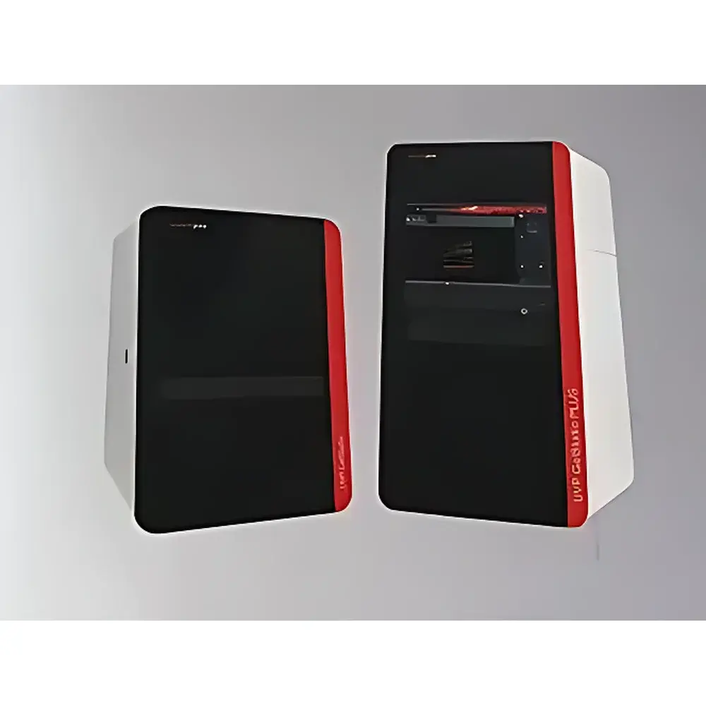
Analytik Jena UVP GelStudio PLUS Multicolor Fluorescence Gel Imaging System
| Brand | Analytik Jena |
|---|---|
| Origin | Germany |
| Model | UVP GelStudio PLUS |
| Instrument Type | Multicolor Fluorescence Gel Imaging System |
| CCD Resolution | 12 MP (Physical Pixels) |
| Bit Depth | 16-bit |
| Light Sources | Cold Cathode UV Transilluminator (302 nm), Reflective White LED, Matrix RGB LED Fluorescence Excitation (470/530/630 nm), Optional Upgrades: Laser NIR (785 nm), Xenon Broadband Illuminator |
| Touchscreen Interface | Integrated 10.1" Capacitive Touch Display |
| Software | UVP VisionWorks LS v5.x (FDA 21 CFR Part 11 Compliant Audit Trail Optional) |
| Compliance | CE, IEC 61010-1, ISO 13485 (Manufacturing Quality System) |
Overview
The Analytik Jena UVP GelStudio PLUS is a high-performance, integrated multicolor fluorescence gel imaging system engineered for rigorous life science laboratories requiring reproducible, quantitative imaging across nucleic acid electrophoresis, protein gels, and whole-organism fluorescence applications. Built upon a modular optical architecture, it employs a scientific-grade 12-megapixel monochrome CCD sensor with true 16-bit dynamic range—enabling linear signal capture across four orders of magnitude (0–65,535 intensity levels). The system utilizes transmissive UV excitation at 302 nm via long-life cold cathode tubes, combined with reflective white LED illumination and a programmable matrix of narrow-band RGB LEDs (470 nm blue, 530 nm green, 630 nm red) for multiplexed fluorophore detection—including SYBR Safe, SYPRO Ruby, Alexa Fluor dyes, and GFP/RFP variants. Its optical path includes fixed f/1.4 lens optics, motorized focus control, and automatic exposure calibration—ensuring consistent pixel-level registration across repeated acquisitions.
Key Features
- 12 MP physical resolution CCD sensor with thermoelectric cooling (−25 °C below ambient), minimizing dark current noise and enabling extended exposure times without saturation.
- Integrated 10.1-inch capacitive touchscreen interface with intuitive workflow-driven UI; also fully compatible with Windows-based remote control via USB or Ethernet.
- Three-stage recessed door mechanism (exclusive to GelStudio PLUS) eliminates protruding hinged doors—reducing footprint and improving ergonomic access in crowded benchtop environments.
- Dedicated matte-finish fluorescent sample tray with chemical-resistant surface and integrated UV-blocking shield—prevents ethidium bromide carryover and minimizes background autofluorescence.
- Modular light engine supports field-upgradable excitation sources: optional 785 nm diode laser for near-infrared detection (e.g., IRDye 800CW) and broadband xenon lamp for continuous-spectrum excitation (250–750 nm).
- Pre-configured acquisition protocols for common applications: DNA EtBr gels, SDS-PAGE Coomassie/Silver staining, Western blots, plant GFP imaging, and viability assays—each optimized for gain, exposure, and binning settings.
Sample Compatibility & Compliance
The GelStudio PLUS accommodates standard electrophoresis formats including mini-gels (up to 15 × 15 cm), midi-gels, blot membranes (PVDF/nitrocellulose), microplates (6–96-well), and whole small-plant specimens (Arabidopsis, seedlings). All illumination modules comply with IEC 62471 photobiological safety standards for UV and visible radiation. The instrument’s electrical design meets CE marking requirements under Directive 2014/30/EU (EMC) and 2014/35/EU (LVD). For regulated environments, VisionWorks LS software optionally enables 21 CFR Part 11-compliant user authentication, electronic signatures, and immutable audit trails—fully supporting GLP and GMP workflows per ISO/IEC 17025 and USP .
Software & Data Management
VisionWorks LS v5.x provides unified acquisition, processing, and analysis in a single application—eliminating cross-platform data migration. Core capabilities include lane and band detection with adaptive thresholding, molecular weight estimation using reference ladders, relative densitometry (with background subtraction and normalization), and multi-channel overlay for co-localization analysis. Export options support TIFF (uncompressed, 16-bit), PNG, JPEG2000, and PDF—with embedded EXIF metadata (exposure time, gain, lens aperture, light source ID). Data files are structured in hierarchical folders compliant with FAIR principles (Findable, Accessible, Interoperable, Reusable), and raw image stacks can be archived directly to network-attached storage (NAS) or LIMS-integrated repositories via configurable SFTP endpoints.
Applications
- Nucleic acid analysis: Quantitative detection of PCR products, restriction digests, and CRISPR editing outcomes on agarose and polyacrylamide gels.
- Protein characterization: High-sensitivity visualization of Coomassie- and silver-stained 1D/2D gels; semi-quantitative comparison of post-translational modifications via fluorescent tags.
- Western blot validation: Chemiluminescent and fluorescent secondary antibody detection with dynamic range sufficient to resolve both low-abundance targets and saturated housekeeping bands.
- Plant phenotyping: Non-invasive longitudinal monitoring of GFP/RFP reporter expression in transgenic Arabidopsis or rice seedlings under controlled white-light or UV-A conditions.
- Cell viability assays: Detection of Calcein AM/EthD-1 dual staining in 96-well plates—leveraging RGB channel separation to distinguish live/dead populations without spectral unmixing artifacts.
FAQ
What is the maximum usable field of view for gel imaging?
The optical system captures a 15 × 15 cm active area at native 12 MP resolution; larger gels require stitching via VisionWorks LS panorama mode.
Can the system perform chemiluminescence imaging?
Yes—when equipped with the optional high-sensitivity low-noise mode and extended exposure (up to 60 min), it supports ECL detection on nitrocellulose and PVDF membranes.
Is remote operation supported over a local network?
Yes—VisionWorks LS includes a dedicated client-server mode allowing simultaneous control from up to four workstations with role-based access permissions.
How is calibration maintained across multiple users and sessions?
The system stores per-user calibration profiles (including flat-field correction, dark-frame subtraction, and lens distortion maps) in encrypted SQLite databases synchronized via domain controller policies.
Does the software support batch processing of large image sets?
Yes—command-line scripting (Python API) and GUI-based batch wizards enable automated lane extraction, background correction, and export to CSV/Excel for downstream statistical analysis in R or GraphPad Prism.
Related Products