ASPEC Flowprobe Flow-Based Microextraction Probe Ion Source
| Brand | ASPEC (Prosolia) |
|---|---|
| Origin | USA |
| Model | Flowprobe |
| Instrument Type | Ambient Ion Source |
| Compatible MS Platforms | Thermo Orbitrap, Bruker timsTOF, SCIEX TripleTOF & QTRAP |
| Sample Interface | Liquid Microjet Surface Sampling Probe (LMJ-SSP) |
| Spatial Resolution | ~630 µm (rapid profiling), down to ~50 µm (high-res DESI-2D integration) |
| Operating Mode | Real-time, continuous-flow in situ microextraction coupled with ESI ionization |
| Optical Guidance | Integrated coaxial camera for real-time probe positioning and surface navigation |
| Application Scope | Label-free molecular imaging of tissues, single-cell metabolomics, spatially resolved lipidomics/peptidomics, forensic toxicology, dried blood spot (DBS) analysis, time-resolved pharmacokinetics |
Overview
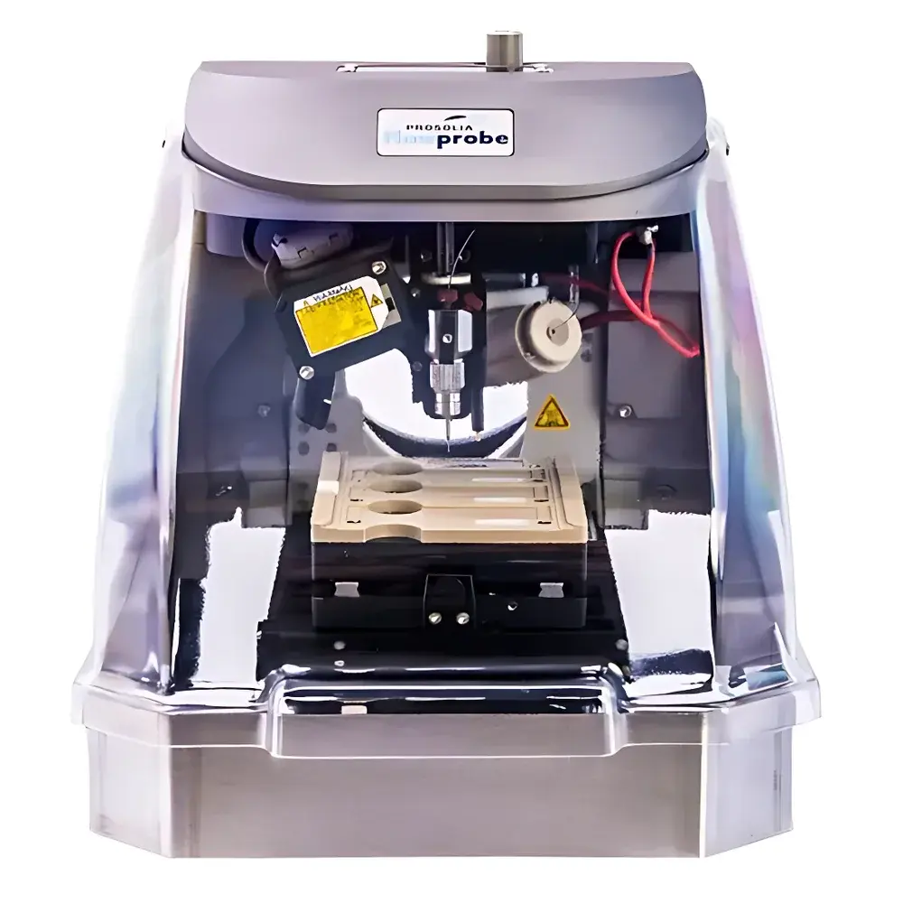
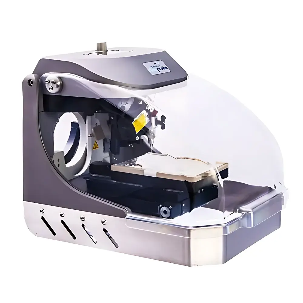
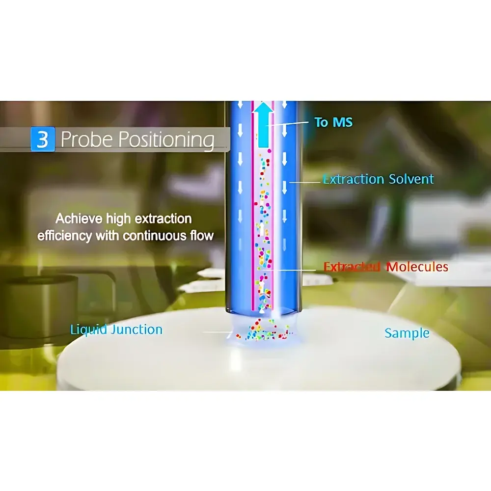
The ASPEC Flowprobe is a next-generation ambient ion source engineered for real-time, in situ microextraction and direct electrospray ionization (ESI) of analytes from planar surfaces. Unlike conventional desorption-based techniques, Flowprobe operates on the Liquid Microjet Surface Sampling Probe (LMJ-SSP) principle—originally developed at Oak Ridge National Laboratory by Dr. Gary Van Berkel—as an evolution of static LESA technology. It employs a coaxial capillary architecture: an outer sheath delivers solvent continuously onto the sample surface to dynamically dissolve and mobilize molecules (e.g., lipids, peptides, drugs, toxins), while an inner capillary simultaneously aspirates the resulting micro-volume extract and transports it directly to an ESI emitter. This closed-loop fluidic interface eliminates offline sample preparation, minimizes analyte degradation, and enables true quantitative potential under controlled flow conditions. The system integrates seamlessly with high-performance mass spectrometers—including Thermo Orbitrap, Bruker timsTOF, and SCIEX QTRAP/TripleTOF platforms—without hardware modification, preserving native instrument sensitivity and mass accuracy.
Key Features
- Coaxial LMJ-SSP architecture enabling simultaneous solvent delivery and extract aspiration at nanoliter-to-microliter flow rates
- Real-time optical guidance via integrated coaxial camera for precise, operator-directed probe positioning over heterogeneous surfaces
- Compatible with both rapid large-area molecular profiling (~630 µm spot size) and high-resolution spatial mapping (~50 µm) when interfaced with DESI-2D motion stages
- No matrix application, no vacuum transfer, and no derivatization required—fully ambient operation at atmospheric pressure
- Robust mechanical design optimized for reproducible probe-to-surface distance control (±2 µm tolerance) across extended scanning sequences
- Modular integration: retrofittable onto existing DESI-2D systems without recalibration or vendor lock-in
Sample Compatibility & Compliance
Flowprobe accommodates a broad range of flat, non-porous to moderately porous substrates—including formalin-fixed paraffin-embedded (FFPE) tissue sections, frozen tissue cryosections, bacterial lawns on agar plates, polymer films, and dried blood spots (DBS). Its solvent-selective extraction capability supports method development per analyte class (e.g., acetonitrile/water for polar metabolites; chloroform/methanol for lipids). The system complies with GLP-aligned workflows: all motion parameters, solvent flow rates, and acquisition timestamps are logged in machine-readable format for audit trail generation. While not a regulated medical device, Flowprobe-generated data meet evidentiary requirements for forensic toxicology (per ASTM E2915-22 guidelines) and support ICH M10-compliant bioanalytical method validation when coupled with appropriate QC standards.
Software & Data Management
Control is managed through Prosolia’s proprietary FlowControl software, which synchronizes probe translation, solvent flow rate (0.1–5.0 µL/min), ESI voltage (±0.5–5.0 kV), and MS acquisition triggers. Raw data export adheres to open mzML 1.1.0 format, ensuring compatibility with downstream tools including SCiLS Lab, MSiReader, and Cardinal for spatial statistics. All acquisition metadata—including camera frames, stage coordinates, and flow sensor readings—are embedded in the mzML header, satisfying 21 CFR Part 11 requirements for electronic records when deployed in validated environments. Batch processing scripts support automated region-of-interest (ROI) extraction and inter-sample normalization using internal standard co-sprayed via auxiliary channel.
Applications
- Label-free spatial metabolomics and lipidomics in tumor margin assessment and cancer subtyping
- Time-resolved drug distribution kinetics in organotypic tissue slices and 3D cell cultures
- High-throughput forensic screening of illicit substances and pharmaceuticals in DBS cards
- In situ characterization of microbial metabolic heterogeneity within biofilms and colony isolates
- Process analytical technology (PAT) monitoring of pharmaceutical coating uniformity and dissolution behavior
- Single-cell peptidomics in neuronal tissue sections under native physiological conditions
FAQ
Is Flowprobe compatible with my existing mass spectrometer?
Yes—Flowprobe interfaces directly with any commercial ESI-compatible mass spectrometer via standard API source ports. No firmware updates or OEM collaboration are required.
What solvents are recommended for lipid extraction?
Chloroform:methanol (2:1 v/v) with 0.1% formic acid is empirically validated for phospholipid and glycerolipid recovery; alternative mixtures can be optimized via FlowControl’s programmable gradient module.
Can Flowprobe be used for quantitative analysis?
Quantitative capability is achievable using isotope-labeled internal standards co-sprayed via the auxiliary solvent channel, with linearity demonstrated across three orders of magnitude (R² > 0.995) in tissue homogenate spike-recovery studies.
Does Flowprobe require special sample preparation?
No matrix, no washing, no vacuum drying—samples are mounted directly on conductive glass slides or aluminum target plates. FFPE sections require only deparaffinization and rehydration prior to analysis.
How is spatial resolution defined and verified?
Resolution is determined by the effective sampling diameter at half-maximum intensity, measured using printed dye arrays and confirmed via edge-spread function analysis per ISO 12233:2017 methodology.


")