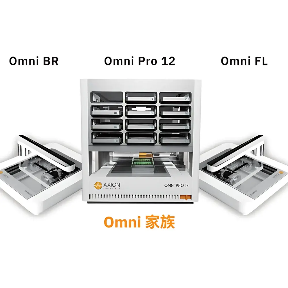
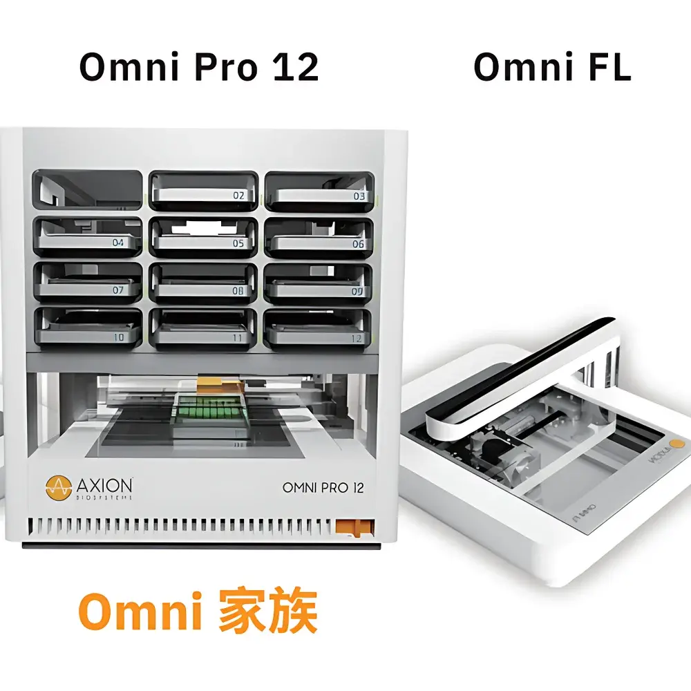
Axion BioSystems Omni BR/FL Live-Cell Real-Time Imaging System for Wound Healing (Scratch Assay) Analysis
| Brand | Axion BioSystems |
|---|---|
| Origin | Netherlands |
| Manufacturer Type | Original Equipment Manufacturer (OEM) |
| Product Category | Imported Instrument |
| Model | Omni |
| Pricing | Available Upon Request |
Overview
The Axion BioSystems Omni BR/FL is a compact, incubator-integrated live-cell imaging workstation engineered for label-free and fluorescence-based dynamic monitoring of adherent mammalian cells in standard multiwell plates and flasks. Operating on the principle of high-resolution brightfield (BR) and epifluorescence (FL) microscopy with motorized stage scanning and automated image stitching, the Omni enables non-invasive, longitudinal acquisition of cellular morphology, confluence, migration, and proliferation under physiologically relevant conditions—without removal from the CO₂ incubator. Its optical architecture employs a fixed LED illumination source positioned above the sample plane and a precision-stepped CMOS sensor mounted beneath the sample stage, enabling consistent axial alignment and minimizing thermal and mechanical perturbation to cultures. Designed for GLP-aligned workflows, the system supports time-lapse imaging at user-defined intervals (from minutes to hours), delivering spatially registered image stacks suitable for quantitative kinetic analysis of cellular behavior—including wound closure kinetics in scratch assays.
Key Features
- Incubator-compatible hardware: Fully operational within standard 5–40 °C, 20–95% RH CO₂ incubators; no external optics or cooling required
- Dual-channel imaging: Simultaneous brightfield and fluorescence (with optional filter cubes) acquisition across all wells
- Full-plate panoramic scanning: Motorized lens movement enables seamless tiling of up to 7850 individual frames per scan, stitched into a single 86 mm × 124 mm composite image (equivalent to full coverage of 6–96-well plates)
- No-plate-handling workflow: Eliminates mechanical stress and microenvironmental disruption caused by plate transfer during imaging
- Cloud-connected data pipeline: Raw TIFF images uploaded automatically to secure CytoSMART Cloud infrastructure; encrypted storage compliant with ISO/IEC 27001 standards
- Zero-calibration design: Factory-aligned optics and embedded auto-focus algorithms ensure reproducible image quality without routine maintenance or user calibration
- Remote operation: Full control via web browser or desktop client—including real-time preview, schedule configuration, and analysis initiation—from any networked device
Sample Compatibility & Compliance
The Omni accommodates all optically transparent cell culture vessels with height ≤55 mm and base thickness ≤1.5 mm, including 6–384-well polystyrene plates, Petri dishes (35–150 mm), T-flasks (T25–T225), and custom microfluidic devices. Its imaging field-of-view (86 × 124 mm) ensures complete coverage of standard 96-well formats and partial-to-full coverage of larger vessels. The system complies with IEC 61000-6-2 (immunity) and IEC 61000-6-3 (emissions) for electromagnetic compatibility. Data handling adheres to ALCOA+ principles (Attributable, Legible, Contemporaneous, Original, Accurate, Complete, Consistent, Enduring, Available); audit trails, electronic signatures, and role-based access control are available through optional CytoSMART Enterprise Software—validated for 21 CFR Part 11 compliance in regulated environments.
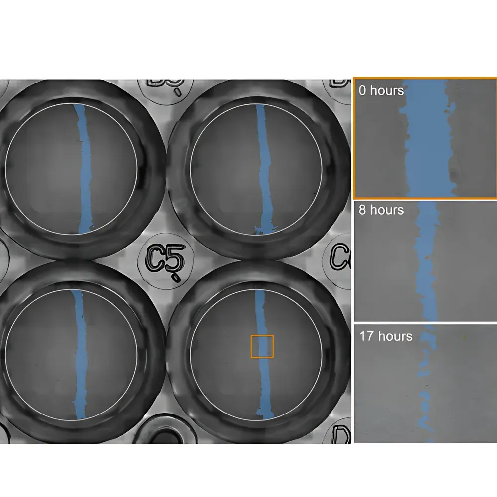
Software & Data Management
CytoSMART Software v5.x provides an intuitive interface for experiment setup, image acquisition scheduling, and algorithm-driven analysis. Pre-installed modules include: (1) Confluence Quantification (brightfield/fluorescence), (2) Wound Healing (Scratch Assay) Analysis, (3) Colony Formation Assay, and (4) Fluorescent Object Counting. All algorithms are validated against ground-truth manual segmentation and report metrics including wound area (µm²), relative wound closure (%), migration velocity (µm/h), and time-to-50%-closure (t₅₀). Raw image data (16-bit TIFF) is exportable for third-party analysis in ImageJ/Fiji, MATLAB, or Python-based pipelines. Cloud-hosted processing supports batch analysis across hundreds of wells, with metadata tagging (e.g., plate ID, treatment group, timepoint) preserved in structured JSON format for integration with LIMS or ELN systems.
Applications
- Wound healing / scratch assay: Quantitative tracking of collective cell migration kinetics in response to pharmacological inhibitors (e.g., blebbistatin, cytochalasin D), growth factors, or genetic perturbations
- Tumor spheroid modeling: Monitoring spheroid compaction, invasion into Matrigel, and drug-induced disintegration over 72–120 h
- Cytotoxicity profiling: Time-resolved assessment of membrane integrity loss (via propidium iodide), metabolic activity (Calcein-AM), or apoptosis markers
- Clonogenic survival: Automated detection and growth curve fitting of colonies formed from single cells post-irradiation or chemotherapeutic exposure
- Immuno-oncology assays: Kinetic evaluation of CAR-T or NK cell-mediated tumor lysis using dual-label fluorescence (e.g., GFP-labeled targets + mCherry-labeled effectors)
- Transfection efficiency monitoring: Temporal quantification of fluorescent protein expression onset and distribution heterogeneity across populations
FAQ
How does the Omni acquire images without disturbing the culture environment?
The system uses a bottom-mounted, motorized CMOS sensor that scans beneath the stationary culture vessel while illumination remains fixed above. This eliminates vibration, temperature fluctuation, and CO₂ loss associated with plate movement.
What image analysis algorithms are included by default?
Standard license includes Brightfield/Fluorescence Confluence, Wound Healing (Scratch Assay), Colony Formation, and Fluorescent Object Counting modules. Additional modules (e.g., Spheroid Area/Volume, Neurite Outgrowth) are available as licensed add-ons.
Can the Omni be integrated into existing laboratory IT infrastructure?
Yes—via RESTful API and SFTP export, raw and processed data can be routed to institutional servers, LIMS, or cloud-based ELNs. On-premise deployment of CytoSMART Enterprise Server is supported for air-gapped networks.
Is validation documentation available for GxP environments?
IQ/OQ protocols, risk assessments, and 21 CFR Part 11 readiness reports are provided with Enterprise Software licensing. Installation Qualification kits include traceable calibration certificates for optical resolution (≥2.5 µm lateral) and illumination uniformity (±5% across FOV).


