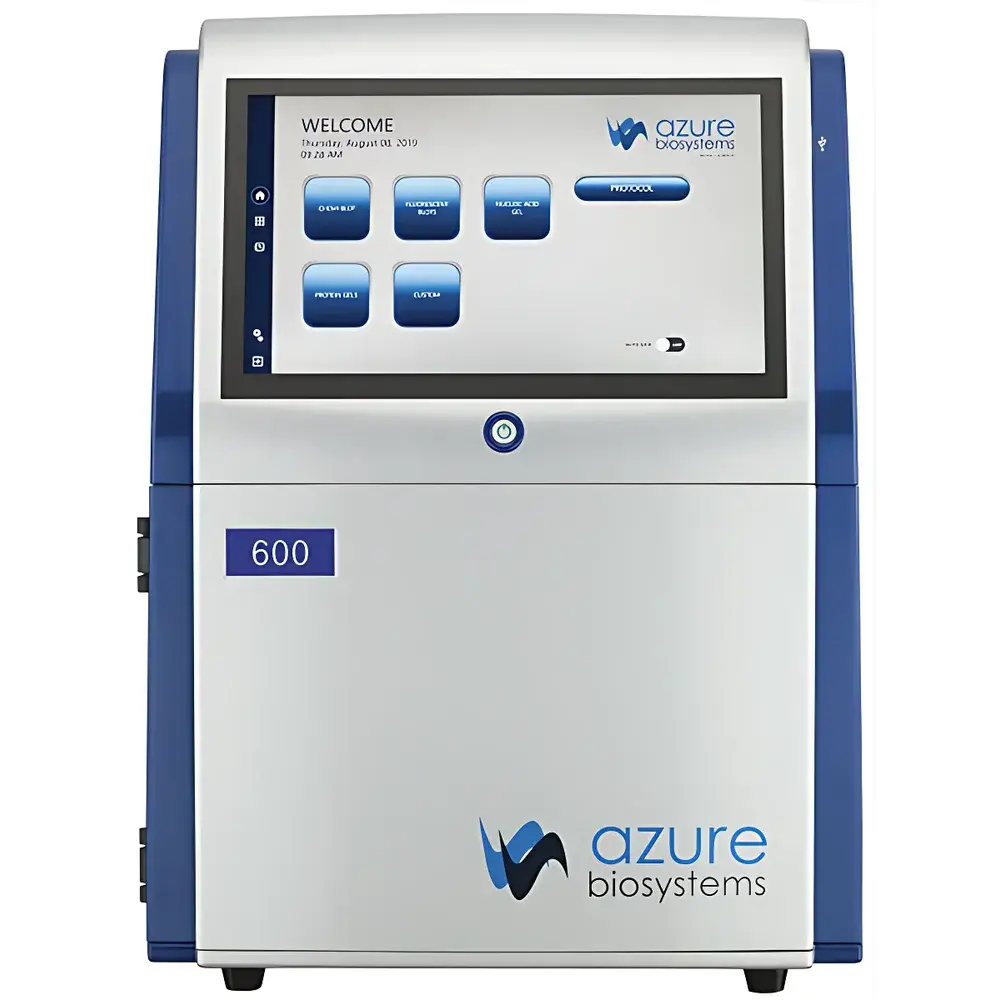
Azure Biosystems Azure 600 Multifunctional Fluorescence Gel Imaging System
| Brand | Azure Biosystems |
|---|---|
| Origin | USA |
| Model | Azure 600 |
| Instrument Type | Multicolor Fluorescence Gel Imaging System |
| CCD Resolution | 9.1 MP |
| Bit Depth | 16-bit |
| Dynamic Range | 4.8 OD |
| CCD Sensor Size | 2/3-inch |
| Detection Sensitivity | fg-level |
| Signal-to-Noise Ratio | High |
| Lens System | Motorized Autofocus |
| Integrated Display | 13.3-inch HD Touchscreen PC |
Overview
The Azure Biosystems Azure 600 Multifunctional Fluorescence Gel Imaging System is an integrated, benchtop digital imaging platform engineered for quantitative molecular biology applications. It employs high-quantum-efficiency CCD detection coupled with multi-spectral excitation and emission filtering to support simultaneous acquisition across visible (RGB), ultraviolet (254 nm and 302 nm), blue-light (470 nm), and dual-laser near-infrared (700 nm and 800 nm) channels. The system operates on the principle of photon capture from fluorophore- or chemiluminescent-labeled biomolecules following electrophoretic separation—enabling precise spatial registration and linear intensity quantification across a 4.8 optical density (OD) dynamic range. Its architecture eliminates external computer dependency by embedding a dedicated 13.3-inch HD touchscreen Windows PC, reducing footprint and minimizing data transfer latency while maintaining full computational control over image acquisition, processing, and export.
Key Features
- Motorized autofocus lens system ensures consistent focal plane alignment across sample types—from thin agarose gels to thick PVDF membranes—without manual intervention.
- Dual UV transilluminators (254 nm and 302 nm) and a high-intensity blue LED (470 nm) enable safe, high-yield excitation of SYBR Safe, Ethidium Bromide, and other nucleic acid dyes.
- Two independently controlled solid-state lasers (685 nm and 785 nm) provide narrow-band excitation for IRDye®, Alexa Fluor®, and LI-COR®-compatible near-infrared probes, minimizing spectral crosstalk in multiplexed western blots.
- 16-bit digitization and 9.1-megapixel CCD sensor deliver high-fidelity spatial resolution (≤ 25 µm pixel size at 1:1 magnification) and low-noise signal capture, critical for detecting faint bands in low-abundance protein or RNA samples.
- Preconfigured and user-defined imaging protocols store exposure time, lens aperture, laser power, and filter selection—ensuring inter-experimental reproducibility and compliance with GLP-aligned laboratory practices.
Sample Compatibility & Compliance
The Azure 600 supports standard electrophoretic formats including mini and midi SDS-PAGE gels, agarose gels (up to 20 × 20 cm), western blot membranes (PVDF and nitrocellulose), and stained tissue sections. It accommodates both wet and dry imaging configurations and integrates seamlessly with common staining reagents (e.g., Coomassie, Silver, SYPRO Ruby) and chemiluminescent substrates (e.g., Luminata Forte, ECL Prime). All image metadata—including timestamp, instrument settings, user ID, and calibration logs—are embedded in TIFF and PNG exports. Software audit trails, electronic signatures, and session locking align with FDA 21 CFR Part 11 requirements for regulated environments, supporting traceability in QC/QA workflows and manuscript submission to journals requiring raw-data transparency (e.g., Nature, Cell, JBC).
Software & Data Management
Acquisition and analysis are managed through AzureSpot™ software—a validated, version-controlled application built on Microsoft .NET Framework. Core capabilities include background subtraction, band intensity normalization (against total lane or reference protein), molecular weight estimation, and multi-channel overlay with false-color assignment. Export options include publication-ready TIFF, PDF, and Excel-compatible CSV files with embedded metadata. Data integrity is preserved via write-once archive mode and optional network storage integration (SMB/NFS). Version history, user access controls, and automated backup scheduling further reinforce data governance in shared core facilities.
Applications
- Quantitative analysis of nucleic acid fragments in agarose and polyacrylamide gels
- Digital documentation and densitometry of Coomassie- and silver-stained protein gels
- Chemiluminescent detection of HRP- and AP-conjugated antibodies in western blotting
- Multiplexed NIR fluorescence imaging for two-color westerns and phosphoprotein profiling
- RGB fluorescence imaging of GFP/RFP-tagged proteins, viability assays (Calcein AM/PI), and histological sections
- Total protein normalization using stain-free or REVERT™ technologies
- Validation of CRISPR editing efficiency via T7E1 or Surveyor assays
FAQ
Does the Azure 600 support remote operation via external computers?
Yes—while fully functional as a standalone unit, it can be controlled remotely via Ethernet or Wi-Fi using the AzureSpot Remote Client, enabling centralized management in multi-user core facilities.
Is the system compatible with third-party analysis software such as ImageJ or Fiji?
All acquired images are saved in standard 16-bit TIFF format with embedded EXIF metadata, ensuring full interoperability with open-source and commercial analysis platforms.
How is calibration maintained across imaging sessions?
The system includes daily auto-calibration routines for lens focus, illumination uniformity, and CCD dark-current subtraction—logged automatically and accessible via the software audit trail.
Can the Azure 600 perform time-lapse or kinetic imaging?
No—it is optimized for endpoint static imaging; kinetic acquisition is not supported due to hardware design focused on maximum sensitivity per exposure rather than frame-rate throughput.
What service and validation documentation is provided?
Each unit ships with Factory Acceptance Test (FAT) reports, IQ/OQ documentation templates, and a Certificate of Conformance compliant with ISO 9001:2015 and IEC 61010-1 safety standards.


