Betop Scientific XPL-1 Polarizing Microscope
| Brand | Betop Scientific |
|---|---|
| Origin | Guangdong, China |
| Manufacturer Type | Direct Manufacturer |
| Country of Origin | China |
| Model | XPL-1 |
| Pricing | Upon Request |
Overview
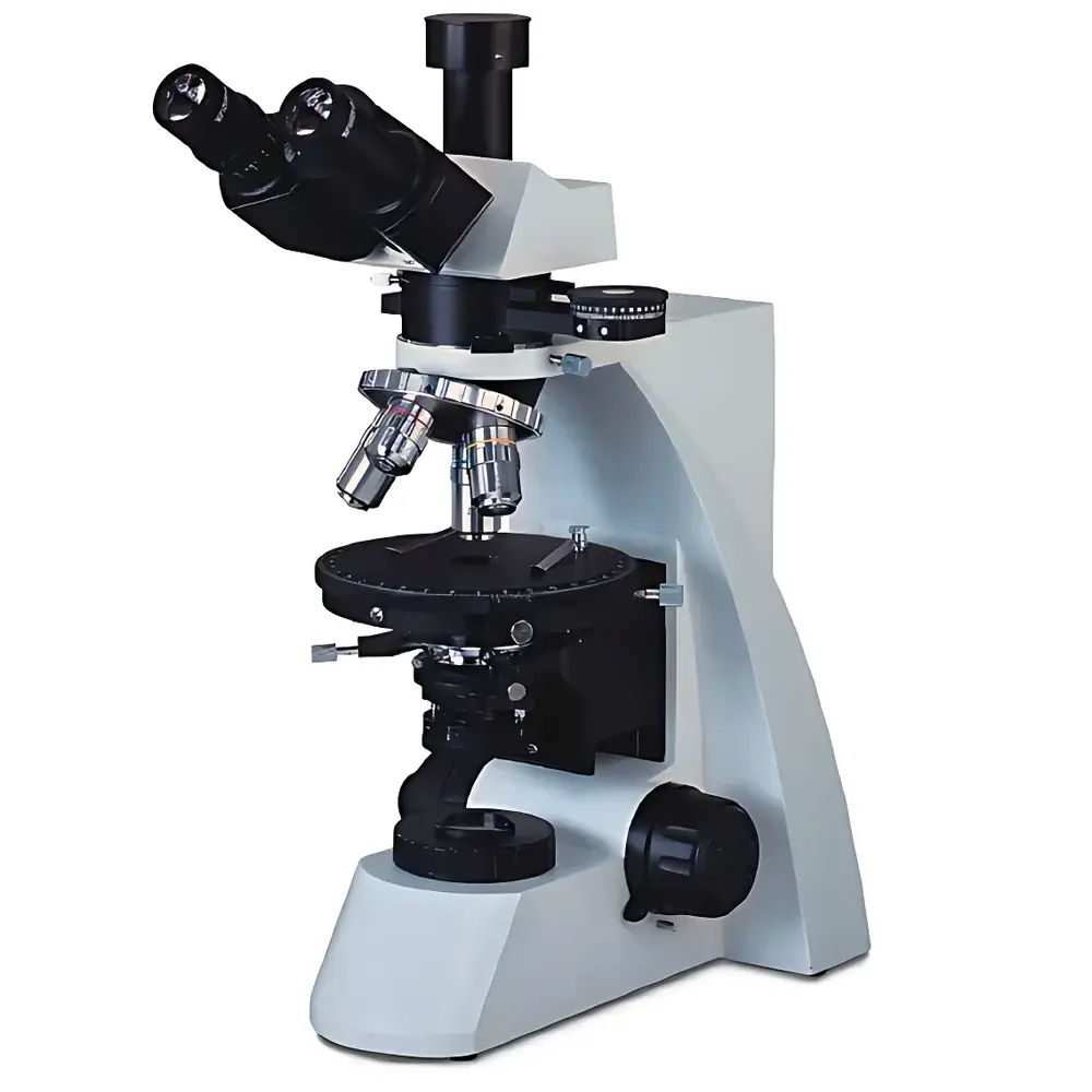
The Betop Scientific XPL-1 Polarizing Microscope is a high-precision transmitted-light optical instrument engineered for quantitative birefringence analysis and anisotropic material characterization. Based on the fundamental principles of polarized light interference—employing orthogonal polarizers, compensators, and convergent illumination—the XPL-1 enables rigorous identification and structural evaluation of crystalline, fibrous, and mineralogical specimens. Its optical architecture adheres to classical polarizing microscopy conventions defined in ASTM E112 (grain size determination), ISO 9276 (particle shape analysis), and USP (microscopic examination of pharmaceutical excipients). Designed for routine use in quality control laboratories, geological survey units, metallurgical R&D facilities, and academic teaching environments, the XPL-1 delivers consistent, repeatable contrast generation under single-polarizer (plane-polarized), crossed-polarizer (extinction and interference figure), and conoscopy (interference pattern) observation modes.
Key Features
- Hinged trinocular head with 30° inclination and adjustable interpupillary distance; supports 100% beam-splitting for simultaneous visual observation and digital imaging via C-mount or DSLR adapters.
- Rotatable analyzer (360° scale with vernier reading to ±12′) with push-in/pull-out mechanism—enabling rapid transition between brightfield and polarized observation without realignment.
- Center-adjustable Bertrand lens with push-in function for precise interference figure acquisition during conoscopy.
- 150 mm diameter rotating mechanical stage with 360° graduated scale (vernier resolution: 6′), centering adjustment, and locking mechanism—optimized for orientation-sensitive crystallographic measurements.
- Integrated λ, λ/4, and quartz wedge compensators—mounted in standardized slots for controlled retardation calibration and sign-of-elongation determination.
- Parfocal, strain-free plan achromatic polarizing objectives (4×, 10×, 40×, 100× oil) featuring anti-reflection coatings, calibrated working distances, and POL designation per DIN 58883 standards.
- Abbe condenser (NA 1.25) with rotating turret for four fixed aperture positions (0°, 90°, 180°, 270°) and iris diaphragm—ensuring optimal Köhler illumination across all magnifications.
Sample Compatibility & Compliance
The XPL-1 accommodates standard 26 mm × 76 mm glass slides and 18 mm circular cover slips. It supports both dry and immersion (Type A cedarwood oil, n = 1.515) mounting media for high-resolution birefringence assessment. Specimen compatibility spans pharmaceutical active ingredients (e.g., polymorphic lactose, mannitol), biological macromolecules (collagen, keratin, starch granules), urological crystals (monosodium urate, calcium oxalate), geological thin sections (quartz, feldspar, mica), and conservation science samples (pigment layers, ground preparations in mural paintings). The system conforms to GLP documentation requirements when paired with audit-trail-capable imaging software and meets mechanical stability criteria outlined in ISO 10934-1 (optical microscopes — terminology and metrological characteristics).
Software & Data Management
While the XPL-1 operates as a standalone optical platform, it integrates seamlessly with third-party image acquisition and analysis suites—including NIS-Elements (Nikon), ZEN Blue (Zeiss), and open-source tools such as ImageJ/Fiji with Polarization Plugin. When equipped with optional USB or HDMI video outputs (up to 10 MP resolution), the microscope supports time-stamped image capture, ROI-based birefringence intensity profiling, and export of TIFF/RAW files compatible with LIMS environments. All digital workflows comply with FDA 21 CFR Part 11 when deployed with validated software packages featuring electronic signatures, user access controls, and immutable audit trails.
Applications
- Pharmaceutical solid-state characterization: polymorph screening, hydrate/anhydrate differentiation, and excipient crystallinity verification per ICH Q5A and Q6A guidelines.
- Clinical crystallography: identification of pathologic urinary crystals and synovial fluid birefringent particles in gout and pseudogout diagnostics.
- Materials science: grain boundary mapping in metallographic etched sections, phase distribution in polymer blends, and stress-induced birefringence in transparent plastics.
- Geosciences: optical indicatrix determination, extinction angle measurement, and optic sign identification in mineral thin sections.
- Cultural heritage analysis: pigment stratigraphy, binder identification, and degradation product localization in historical painting cross-sections.
FAQ
What types of compensation plates are compatible with the XPL-1?
The microscope accepts standard λ (530 nm), λ/4 (quarter-wave), and quartz wedge compensators—each inserted into dedicated slots beneath the objective nosepiece for precise retardation control.
Is the XPL-1 suitable for quantitative retardation measurement?
Yes—when used with calibrated compensators and a calibrated analyzer scale, the system supports semi-quantitative retardation estimation using the Sénarmont method or Michel-Lévy chart correlation.
Can the XPL-1 be upgraded with motorized components?
No native motorization is available; however, third-party motorized stages and focus drives can be retrofitted with mechanical interface adaptation.
Does the XPL-1 meet ISO/IEC 17025 requirements for accredited testing labs?
As an optical instrument, it serves as a validated measurement tool within accredited workflows when accompanied by documented calibration records, operator training logs, and traceable reference standards.
What is the maximum usable magnification under oil immersion?
With the 100×/1.25 POL objective and Type A immersion oil, the theoretical maximum useful magnification is 1250×, consistent with Abbe diffraction limits for visible light (λ ≈ 550 nm).


