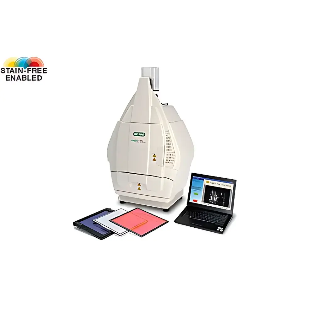
Bio-Rad ChemiDoc XRS+ Chemiluminescence Gel Imaging System
| Brand | Bio-Rad |
|---|---|
| Origin | USA |
| Model | ChemiDoc XRS+ |
| Instrument Type | Chemiluminescence Gel Imaging System |
| CCD Resolution | 4 MP |
| Bit Depth | 16-bit |
| Dynamic Range | 0–4.8 OD |
| CCD Sensor Size | 1-inch |
| Detection Sensitivity | pg–fg level for proteins |
| Signal-to-Noise Ratio | >56 dB |
| Lens | Motorized Zoom Lens |
| Imaging Modes | Chemiluminescence, Fluorescence (single/dual-color), Colorimetric, Densitometry |
Overview
The Bio-Rad ChemiDoc XRS+ Chemiluminescence Gel Imaging System is a high-performance, fully integrated digital imaging platform engineered for quantitative analysis of nucleic acid gels, protein gels, and blot membranes. It employs a thermoelectrically cooled 4-megapixel scientific-grade CCD detector with 16-bit digitization and a calibrated optical path to deliver exceptional sensitivity and reproducibility across chemiluminescent, fluorescent, and colorimetric detection modalities. The system operates on the principle of photon capture under controlled darkroom conditions, where emitted light from labeled biomolecules—whether generated via enzyme-substrate reactions (e.g., HRP-luminol), direct fluorophore excitation, or chromogenic development—is focused onto the CCD sensor through a motorized zoom lens assembly. Its 0–4.8 optical density (OD) dynamic range enables linear quantification over four orders of magnitude, supporting rigorous comparative analysis in applications ranging from Western blot validation to multiplexed fluorescence-based protein profiling.
Key Features
- Thermoelectrically cooled 4 MP CCD sensor (1-inch format) minimizing thermal noise and enabling long-exposure acquisition without signal degradation
- Motorized zoom lens with auto-focus and preset magnification calibration for consistent field-of-view reproducibility across users and sessions
- Integrated white-light transilluminator and dual-band LED excitation sources (470 nm & 530 nm) for simultaneous single- or dual-color fluorescence imaging
- High-sensitivity chemiluminescence mode optimized for low-abundance targets, achieving detection limits in the pg–fg range for immunodetected proteins
- Onboard image acquisition software with real-time histogram feedback, exposure optimization algorithms, and automatic background subtraction
- Robust mechanical shutter and light-tight enclosure certified for ambient-light-free operation during chemiluminescent acquisitions
Sample Compatibility & Compliance
The ChemiDoc XRS+ supports standardized electrophoretic and blotting formats including mini-gels (up to 10 × 10 cm), standard gels (up to 16 × 20 cm), and transfer membranes (PVDF, nitrocellulose, nylon). It is validated for use with common nucleic acid stains (Ethidium Bromide, SYBR® Green I/II, SYBR® Safe, GelRed™, GelGreen™), protein stains (Coomassie Brilliant Blue R-250, Silver Nitrate, SYPRO Ruby, Krypton, Flamingo), and immunodetection reagents (HRP- and AP-conjugated secondary antibodies, Qdot® nanocrystals, Alexa Fluor® dyes, DyLight® conjugates). The system complies with GLP/GMP documentation requirements through audit-trail-enabled software logging, user-access controls, and exportable metadata (including exposure time, gain, lens position, and calibration date). Image files are saved in TIFF format with embedded EXIF metadata, facilitating traceability in regulated environments aligned with FDA 21 CFR Part 11 and ISO/IEC 17025 guidelines.
Software & Data Management
Acquisition and analysis are performed using Bio-Rad’s Image Lab Touch Software—a touch-optimized, modular application supporting both standalone and network-deployed configurations. The software provides automated lane and band detection, molecular weight estimation, relative quantification (normalized to loading controls or total protein), and statistical comparison across replicates. All processing steps—including background correction, smoothing, thresholding, and densitometric integration—are non-destructive and fully reversible. Raw image data and processing history are stored in a relational database with timestamped operator logs. Export options include publication-ready PNG/PDF, spreadsheet-compatible CSV, and MIAME-compliant XML for integration with LIMS or ELN systems. Software updates follow a documented change control process compliant with laboratory quality management systems.
Applications
- Quantitative Western blotting with chemiluminescent substrates (e.g., Clarity™ ECL, Luminata™ Crescendo)
- Dual-color fluorescence detection of co-immunoprecipitated complexes or post-translational modifications
- Nucleic acid gel documentation with UV-free, non-mutagenic stains for routine PCR and restriction digest analysis
- Protein expression profiling via Coomassie- or silver-stained 2D gels with spot matching and volume ratio analysis
- Validation of CRISPR/Cas9 editing efficiency using T7E1 or Surveyor nuclease assays
- Quality control of recombinant protein purification batches through SDS-PAGE densitometry
FAQ
What is the maximum gel/membrane size supported by the ChemiDoc XRS+?
The system accommodates samples up to 16 cm × 20 cm, compatible with standard Mini-PROTEAN® and Criterion™ gel formats as well as full-size transfer membranes.
Does the ChemiDoc XRS+ support time-lapse chemiluminescence imaging?
Yes—the system allows programmable multi-frame acquisition with variable exposure times, enabling kinetic monitoring of luminescent signal decay or substrate reaction progression.
Can Image Lab Touch Software be validated for use in regulated pharmaceutical labs?
Yes—Bio-Rad provides IQ/OQ documentation packages, electronic signature templates, and 21 CFR Part 11 configuration guidance to support validation under GxP frameworks.
Is calibration required before each use?
While daily calibration is not mandatory, Bio-Rad recommends performing flat-field and dark-current calibration at least once per week or after major environmental changes (e.g., temperature shifts >5°C) to maintain quantitative fidelity.
How does the system handle autofluorescence from PVDF membranes?
The motorized zoom lens includes spectral filtering options; combined with software-based background modeling and subtraction algorithms, it effectively suppresses membrane autofluorescence in both single- and dual-channel modes.


