

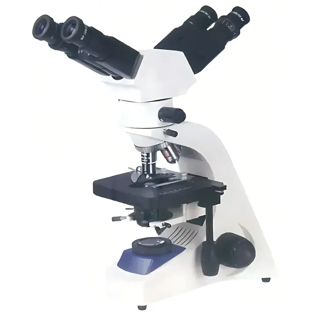
BM-18F2 Dual-Head Teaching Biological Microscope (BM Brand)
| Brand | BM |
|---|---|
| Origin | Shanghai, China |
| Model | BM-18F2 |
| Microscope Type | Inverted Teaching Microscope |
| Eyepiece Configuration | Binocular |
| Optical System | Infinity-Corrected |
| Illumination | 6V/20W Halogen with Köhler Illumination |
| Objective Lenses | 4×, 10×, 40×(S), 100×(S) Oil Immersion (Achromatic Infinity-Corrected) |
| Eyepieces | Widefield 10×/22 mm |
| Stage | Double-layer Mechanical Stage (180 × 150 mm |
| Condenser | NA 1.25 Abbe Condenser with Iris Diaphragm and Filter Holder |
| Focus Mechanism | Coaxial Coarse/Fine Focus (Fine Focus Graduation: 2 µm) |
| Interpupillary Adjustment Range | 55–75 mm |
Overview
The BM-18F2 Dual-Head Teaching Biological Microscope is an engineered solution for collaborative microscopy in academic laboratories, clinical training environments, and biomedical research settings. Designed around an infinity-corrected optical architecture, it delivers high-fidelity, low-aberration imaging across the full magnification range (40×–1000×). Its inverted configuration enables stable observation of live specimens in culture dishes, flasks, or Petri plates—particularly advantageous for cell biology, tissue culture monitoring, and developmental studies. The integrated LED pointer indicator operates optically within the shared optical path, allowing instructors to annotate real-time microscopic fields without obstructing the specimen or requiring external projection systems. This feature supports pedagogical best practices by enabling synchronized visual guidance during group instruction, peer review, or diagnostic case discussions.
Key Features
- Infinity-corrected achromatic objective lens set (4×, 10×, 40× spring-loaded, 100× oil immersion) optimized for consistent resolution and flat-field performance.
- Dual-head binocular observation tube with adjustable interpupillary distance (55–75 mm), accommodating diverse user anthropometrics and supporting simultaneous viewing by instructor and student.
- LED-based optical pointer system: arrow-shaped cursor projected directly into the image plane; position is manually adjustable via precision-mounted control knobs; brightness independently regulated to maintain contrast integrity.
- Köhler illumination system with 6V/20W halogen lamp ensures uniform, glare-free field illumination; voltage compatibility (110V/220V) facilitates international deployment without transformer dependency.
- NA 1.25 Abbe condenser with centerable iris diaphragm and integrated filter holder supports critical aperture control for optimal contrast and resolution in both brightfield and phase contrast applications (when equipped with optional phase components).
- Coaxial coarse/fine focusing mechanism with 2 µm fine-focus graduation enables precise Z-axis positioning essential for serial section analysis, focus stacking, and live-cell time-lapse documentation.
Sample Compatibility & Compliance
The BM-18F2 accommodates standard 35 mm, 60 mm, and 100 mm Petri dishes; T25/T75 tissue culture flasks; multi-well plates (6–96-well); and glass-bottom dishes up to 1.5 mm thickness. Its stage clearance and objective working distances (e.g., 0.15 mm for 100× oil) are calibrated per ISO 8578:2017 (Microscopes — Requirements for mechanical and optical characteristics). While not certified for GLP or GMP-regulated production environments, its optical traceability, repeatable focusing, and standardized mounting interfaces (RMS thread, 23.2 mm eyepiece tubes) support audit-ready documentation workflows in educational QA programs and preclinical research labs operating under institutional biosafety and equipment validation protocols.
Software & Data Management
The BM-18F2 functions as a standalone optical platform compatible with third-party digital imaging solutions. When paired with C-mount adapters and CMOS sensors (e.g., 5 MP or higher resolution), it supports time-stamped image capture, multi-channel overlay, and basic morphometric analysis via open-standard software suites such as ImageJ/Fiji, NIS-Elements (Nikon), or ZEN (Zeiss). Though no proprietary acquisition software is bundled, the microscope’s stable mechanical design and vibration-damped base ensure high reproducibility for longitudinal image datasets—critical for student lab reports, publication-grade figure generation, and internal method validation. All optical adjustments (focus, condenser centering, pointer alignment) are mechanically retained, eliminating recalibration drift between sessions.
Applications
- Undergraduate and graduate-level histology, cytology, and microbiology instruction, where real-time annotation enhances conceptual anchoring during slide interpretation.
- Clinical pathology training: simultaneous observation of stained tissue sections or peripheral blood smears with instructor-guided identification of diagnostic features (e.g., mitotic figures, nuclear atypia, parasite morphology).
- Cell culture QC: routine assessment of confluency, morphology, contamination (yeast, bacteria, mycoplasma), and transfection efficiency in adherent and suspension lines.
- Live-cell imaging workflows: monitoring wound-healing assays, phagocytosis, or neurite outgrowth under ambient CO₂ conditions when used with environmental chamber accessories.
- Comparative morphology studies in botany and zoology labs, leveraging the widefield 22 mm field number for rapid scanning of whole-mount preparations.
FAQ
Is the BM-18F2 compatible with digital camera integration?
Yes—the microscope includes a standard 23.2 mm photo tube port and accepts C-mount adapters for direct coupling to scientific CMOS or CCD cameras.
Can the LED pointer be disabled during independent student use?
Yes—the pointer circuit includes an on/off switch and independent brightness control, allowing full optical path transparency when annotation is not required.
What immersion media are supported by the 100× objective?
The 100× S objective is designed for Type A immersion oil (nD = 1.515–1.520); silicone oil (nD = 1.40) compatibility is not specified and not recommended.
Does the microscope meet CE or FDA regulatory requirements?
As a Class I non-active laboratory instrument intended for teaching and research—not clinical diagnostics—it complies with general safety standards (IEC 61010-1) but does not carry CE marking for medical device use or FDA 510(k) clearance.
How is Köhler illumination aligned on this model?
Alignment follows standard procedure: center the condenser using adjustment screws, close the field diaphragm, focus its edge using the condenser height knob, then open it to just beyond the field of view while adjusting the aperture diaphragm for optimal contrast/resolution balance.


