

BM-18F3 Trinocular Teaching Biological Microscope by BM (Shanghai)
| Brand | BM |
|---|---|
| Origin | Shanghai, China |
| Model | BM-18F3 |
| Microscope Type | Upright Biological Microscope |
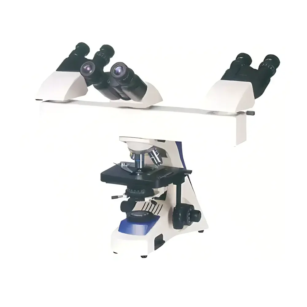
| Observation Head | Hinged Trinocular Head with Interpupillary Adjustment (55–75 mm) |
| Eyepieces | Widefield 10×/22 mm |
| Objectives | Infinity-Corrected Achromatic Objectives — 4×, 10×, 40×(S), 100×(S) Oil Immersion |
| Stage | Dual-layer Mechanical Stage (180 × 150 mm) |
| Travel Range | 75 × 50 mm |
| Condenser | Abbe Condenser, NA 1.25, with Iris Diaphragm and Filter Holder |
| Focus Mechanism | Coaxial Coarse/Fine Focus |
| Fine Focus Graduation | 2 µm |
| Illumination | 6 V / 20 W Halogen Lamp (110 V / 220 V Compatible) |
| LED Pointer System | Adjustable Brightness, Arrow-Shaped Cursor, Fully Movable Within Field of View |
Overview
The BM-18F3 Trinocular Teaching Biological Microscope is an upright, infinity-corrected optical instrument engineered for collaborative life science education, clinical demonstration, and routine histological examination. Designed around a robust mechanical platform and precision optical architecture, it supports simultaneous observation by up to three users—two via the dual eyepiece ports and one via a dedicated trinocular phototube output—enabling real-time instructor-led guidance, peer discussion, and digital image capture. Its optical path integrates Köhler illumination for uniform field brightness and optimal contrast, while the NA 1.25 Abbe condenser ensures efficient light collection across all magnifications, including oil immersion at 100×. The inclusion of an integrated, user-controllable LED pointer system—featuring an arrow-shaped cursor with independent brightness adjustment and full XY mobility within the visual field—facilitates precise annotation during live demonstrations without requiring external annotation software or screen overlays.
Key Features
- Trinocular observation head with adjustable interpupillary distance (55–75 mm) and fixed 30° inclination for ergonomic viewing across diverse user heights and postures.
- Infinity-corrected achromatic objective set (4×, 10×, 40× spring-loaded, 100× oil immersion) delivering consistent resolution and minimal chromatic aberration across the full magnification range.
- Dual-layer mechanical stage with calibrated movement (75 × 50 mm travel) and vernier scale for repeatable specimen repositioning—critical for comparative analysis and serial section alignment.
- Coaxial coarse/fine focus mechanism with 2 µm fine-focus graduation and tension-adjustable rack-and-pinion drive, ensuring stable, vibration-damped focusing at high magnifications.
- LED pointer subsystem mounted in the intermediate optical path: cursor position is optically conjugated to the specimen plane, enabling accurate spatial referencing without parallax error; brightness and on/off state are independently controllable via front-panel button.
- Halogen illumination system with voltage auto-sensing (110 V / 220 V), dimmable output, and built-in heat-absorbing filter to protect specimens from thermal degradation during prolonged observation.
Sample Compatibility & Compliance
The BM-18F3 accommodates standard glass microscope slides (76 × 26 mm) and coverslips (No. 1.5, 0.17 mm thickness), supporting both stained and unstained biological preparations—including hematoxylin-eosin (H&E), Gram-stained smears, blood films, and live tissue sections in aqueous mounting media. Its optical design conforms to ISO 8578:2017 (Microscopes — Requirements for Biological Microscopes) and supports compliance with GLP documentation workflows when paired with validated digital imaging systems. While not certified for IVD or diagnostic use per FDA 21 CFR Part 809, its stable illumination, reproducible focus repeatability, and standardized mechanical interfaces align with internal QC protocols in academic core facilities and teaching laboratories operating under ISO/IEC 17025-aligned quality management systems.
Software & Data Management
The trinocular port accepts C-mount adapters (0.5× or 1× relay lenses) compatible with industry-standard CMOS/CCD cameras (e.g., Thorlabs, AmScope, or Basler models). When integrated with third-party acquisition software such as NIS-Elements (Nikon), ZEN (Zeiss), or open-source platforms like MicroManager, the BM-18F3 supports time-lapse imaging, multi-channel fluorescence registration (when equipped with appropriate filter cubes), and metadata-tagged image export (TIFF, PNG, ND2). The LED pointer position is not digitally encoded but remains optically persistent in captured frames—enabling manual annotation traceability in audit-ready reports. All hardware controls (illumination intensity, pointer activation) are fully manual, eliminating firmware dependencies and ensuring long-term operational consistency in regulated environments where software validation is constrained.
Applications
- Undergraduate and graduate histology, cytology, and parasitology laboratories—where instructor-guided morphological assessment of tissue architecture, cellular organelles, and microbial morphology is essential.
- Clinical pathology training modules involving differential white blood cell identification, urine sediment analysis, and fungal element recognition in direct wet mounts.
- Research support in developmental biology labs performing low-magnification embryo staging and organ rudiment mapping using transmitted-light brightfield.
- Continuing medical education (CME) workshops requiring real-time consensus review of biopsy specimens among multidisciplinary teams.
- Quality assurance checks in biomanufacturing settings for visual verification of cell monolayer confluence prior to passaging or harvest.
FAQ
Is the BM-18F3 compatible with digital camera integration?
Yes—the trinocular port accepts standard C-mount adapters for seamless coupling with scientific-grade cameras. Relay lens magnification must be selected based on sensor size to preserve field-of-view coverage.
Can the LED pointer be used with oil immersion objectives?
Yes—the pointer remains in focus and parallax-free across all objective magnifications, including 100× oil immersion, due to its placement in the infinity space of the optical train.
Does the microscope support phase contrast or fluorescence observation?
The base configuration is optimized for brightfield transmission microscopy. Phase contrast requires optional slider-based annuli and phase objectives; fluorescence capability necessitates dedicated filter cubes, excitation sources, and barrier filters—not included in the standard BM-18F3 package.
What maintenance is required for long-term optical stability?
Annual cleaning of optical surfaces with lens-grade solvents and lint-free wipes is recommended. Halogen lamp replacement should follow manufacturer-rated lifetime (typically 1,000 hours); avoid touching quartz envelopes with bare fingers.
Is service and calibration support available outside mainland China?
BM provides technical documentation and spare parts lists globally. Local authorized distributors offer field service contracts; calibration of mechanical components (stage travel, focus graduation) may be performed in-house using NIST-traceable micrometers and stage micrometers.


