BM-Beam BM-40YD Inverted Fluorescence Microscope
| Brand | BM-Beam |
|---|---|
| Origin | Shanghai, China |
| Model | BM-40YD |
| Instrument Type | Inverted Fluorescence Microscope |
| Research Grade Classification | Yes |
| Fluorescence Filter Cubes | UV1 (385 nm center), B1 (470 nm center), G1 (560 nm center), R1 (630 nm center, optional) |
| Light Source | LED-based, 385 nm / 470 nm / 560 nm triple-channel excitation |
| Control Interface | PC software-controlled fluorescence filter turret and illumination |
| Focusing Mechanism | Coaxial coarse/fine manual focus with 0.002 mm fine adjustment resolution |
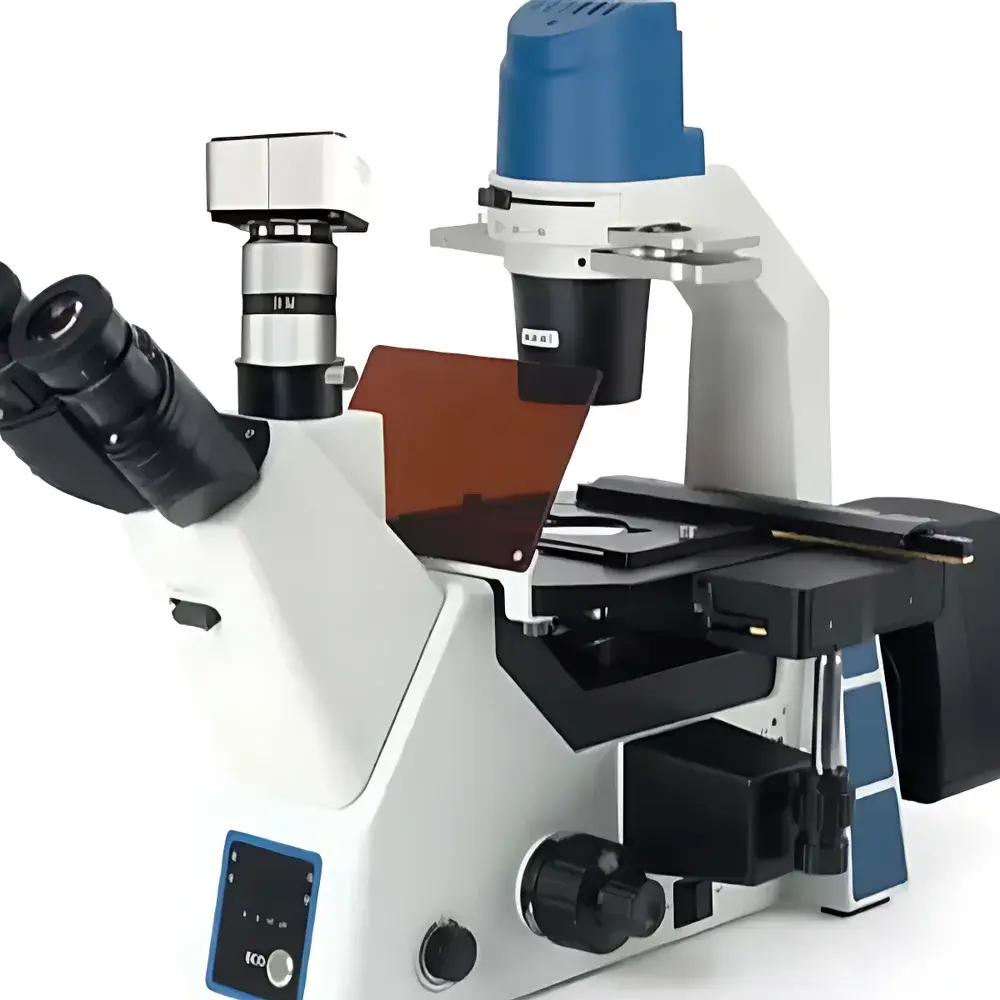
| Condenser | NA 0.3, long working distance (WD = 72 mm), removable, phase-adjustable slider with dual centering knobs |
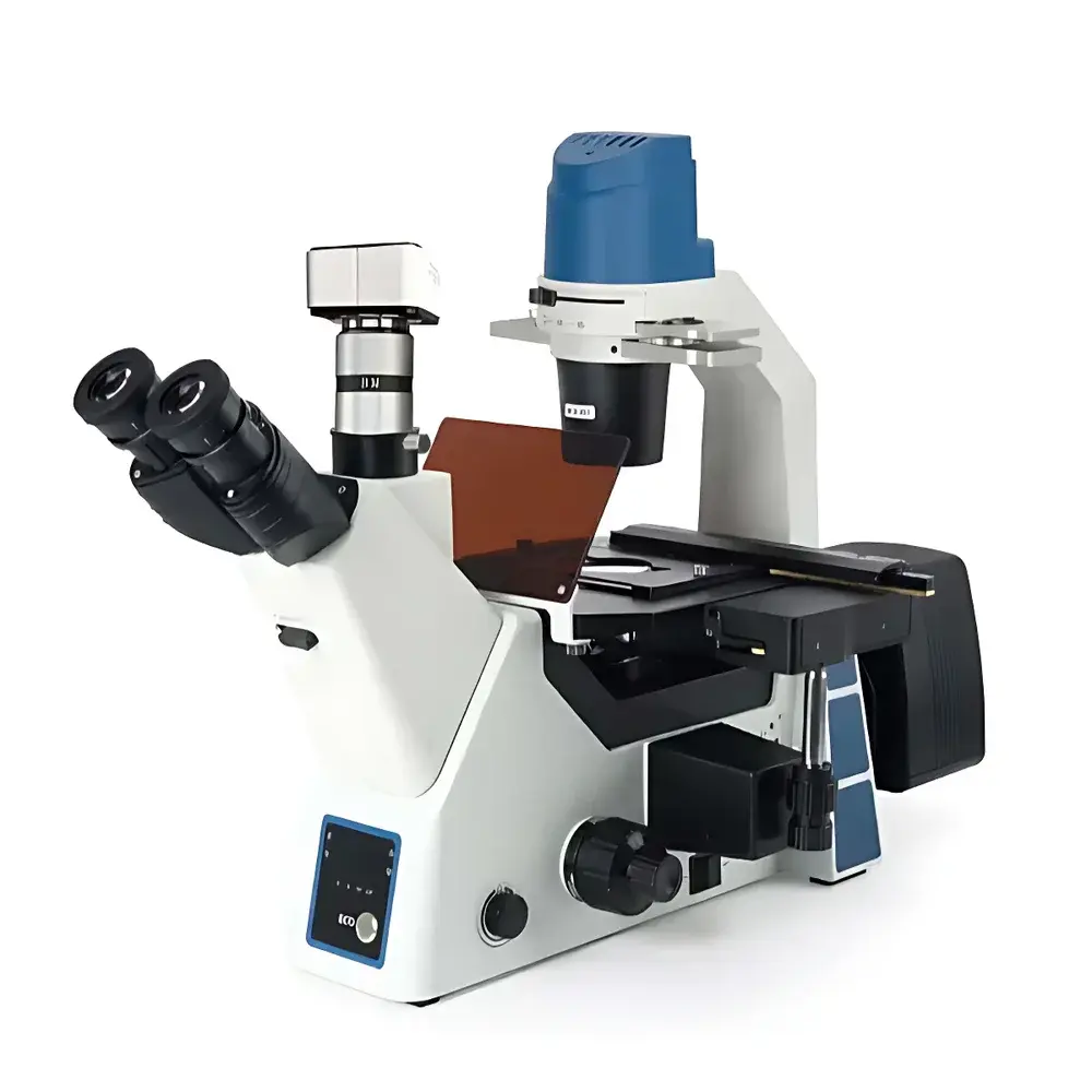
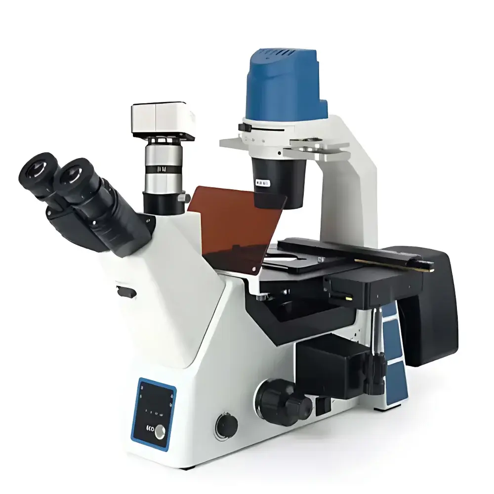
| Observation Head | 45° inclined hinge-type trinocular, 360° rotatable, interpupillary distance 50–75 mm, dual-mode beam splitter (100% eyepiece / 100% camera) |
| Stage | Three-layer mechanical stage, 215 × 250 mm platform, 120 × 80 mm travel range |
| Objective Lenses | Infinity-corrected semi-apochromatic plan fluor objectives (4×/0.13, WD 18.5 mm |
| Optical System | Infinity-corrected semi-plan achromat optical design |
| Illumination | Adjustable warm/cool white transmitted LED, independent intensity control per channel, IR motion sensing, LED fluorescence status indicator |
| Fluorescence Light Engine | Four-position motorized filter turret with servo-driven gear transmission, integrated LED driver for simultaneous or sequential excitation control |
Overview
The BM-Beam BM-40YD Inverted Fluorescence Microscope is a research-grade, modular inverted imaging platform engineered for high-reproducibility live-cell observation, fixed-tissue fluorescence analysis, and multi-modal quantitative microscopy in academic, pharmaceutical, and biotechnology laboratories. Its inverted configuration enables stable long-term imaging of adherent mammalian cells, organoids, and microfluidic culture systems without physical interference from objective lenses—critical for time-lapse experiments, electrophysiology integration, and environmental chamber compatibility. The system employs an infinity-corrected semi-apochromatic optical path optimized for minimal chromatic aberration across visible and near-UV wavelengths, ensuring faithful spectral fidelity during multi-color fluorescence acquisition. Designed to meet the operational rigor of GLP-compliant workflows, the BM-40YD supports full audit-trail logging via its PC-integrated control architecture and maintains alignment stability under continuous thermal load—key attributes for longitudinal studies requiring pixel-level registration consistency.
Key Features
- Inverted ergonomic chassis with reinforced base and vibration-damped stage mount for enhanced mechanical stability during extended acquisitions
- Motorized four-position fluorescence filter turret with servo-controlled positioning and TTL synchronization for precise excitation timing
- Dual-path trinocular head with 100% light routing options (eyepiece-only, camera-only, or 50:50 split), supporting simultaneous visual monitoring and digital capture
- Three-layer mechanical stage featuring micrometer-scale vernier scales and anti-backlash gearing for sub-micron reproducible XY repositioning
- Long-working-distance condenser (NA 0.3, WD 72 mm) with detachable phase slider and dual-axis centering mechanism—enabling rapid transitions between phase contrast and fluorescence modes
- Triple-wavelength LED excitation engine (385 nm, 470 nm, 560 nm) with independent current regulation, thermal feedback control, and <1% intensity drift over 4-hour operation
- Coaxial coarse/fine focusing assembly with 9 mm total travel (6.5 mm upward, 2.5 mm downward), 0.002 mm fine-step resolution, and slip-resistant torque adjustment
Sample Compatibility & Compliance
The BM-40YD accommodates standard 35 mm, 60 mm, and 100 mm Petri dishes; multi-well plates (6–96-well); glass-bottom culture dishes (thickness #1.5 coverslip equivalent); and custom microfluidic devices up to 25 mm height clearance. Its open-stage architecture allows seamless integration with temperature-controlled stages, CO2 incubators, and patch-clamp rigs. The microscope conforms to IEC 61000-6-3 (EMC emissions) and IEC 61000-6-2 (immunity) standards. While not classified as a medical device (non-CE-IVD, non-FDA 510(k)), it supports documentation protocols aligned with ISO/IEC 17025 calibration traceability requirements and provides configurable metadata embedding (EXIF + custom tags) for regulatory-compliant image archiving under 21 CFR Part 11–enabled software environments.
Software & Data Management
Control and acquisition are managed through BM-Beam’s proprietary MicroVision Pro software suite (Windows 10/11, 64-bit), which delivers synchronized hardware orchestration—including motorized filter selection, LED intensity ramping, Z-stack sequencing, and time-lapse scheduling. The software supports TIFF, OME-TIFF, and ND2 export formats with embedded spatial calibration, exposure metadata, and fluorescence channel labeling. Geometric measurement tools include calibrated distance, angle, area, circularity, linearity, and polygonal ROI quantification—with batch-processing capability across multi-field datasets. Raw image data is stored with SHA-256 checksums; acquisition logs record timestamp, user ID, objective ID, filter position, exposure parameters, and environmental sensor inputs (if external sensors are connected). Optional API access enables integration with LabVantage, Benchling, or custom LIMS platforms via RESTful endpoints.
Applications
- Live-cell calcium imaging using Fluo-4 or GCaMP reporters under 470 nm excitation
- Subcellular localization studies (e.g., GFP-tagged organelles, mCherry-labeled cytoskeletal elements)
- Immunofluorescence screening of tissue sections and cell monolayers with DAPI/FITC/TRITC/Cy5 multiplexing
- Phase contrast–fluorescence correlation imaging for label-free morphological validation alongside molecular signal detection
- Quantitative wound-healing assays with automated edge-detection and migration velocity profiling
- Stem cell differentiation monitoring via nuclear morphology (DAPI) and lineage-specific reporter expression
FAQ
Is the BM-40YD compatible with third-party cameras?
Yes—the microscope features a standardized C-mount interface (1× magnification) and supports industry-standard USB3/PCIe cameras via SDK integration. Full trigger and gain control are available through MicroVision Pro’s hardware abstraction layer.
Can the R1 filter cube (630 nm) be installed post-purchase?
Yes—the four-slot filter turret accepts field-upgradable cubes; the R1 set is sold separately and includes matched dichroic and emission filters calibrated for Cy5 and similar dyes.
Does the system support autofocus for time-lapse experiments?
No—autofocus is not natively embedded; however, the fine-focus mechanism is programmable via software API, enabling integration with external Z-drives or piezo actuators for closed-loop focus stabilization.
What is the maximum sample thickness supported with the 40× objective?
With the 40×/0.65 NA objective and standard #1.5 coverslip, the effective working distance is 1.6 mm; total accessible sample height—including dish bottom and medium—is ≤3.2 mm when using the lowest condenser position.
Are maintenance logs and calibration certificates provided?
Yes—each unit ships with a factory calibration report (including Köhler alignment verification, filter transmission spectra, and LED output stability test data) and a logbook template compliant with ISO/IEC 17025 documentation practices.


