

BM Optical XSP-BM-17A Upright Biological Microscope
| Brand | BM Optical |
|---|---|
| Origin | Shanghai, China |
| Microscope Type | Upright |
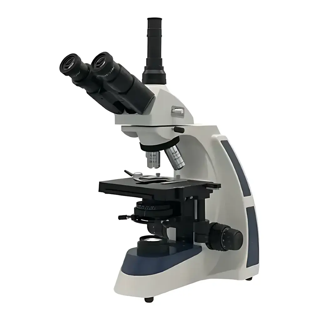
| Configuration | Trinocular |
| Optical Design | UIS Infinity-Corrected System |
| Eyepieces | Widefield 10×/Φ22 mm (standard), optional 16×/Φ15 mm |
| Objectives | Infinity-Corrected Achromatic, 4×/0.10, 10×/0.25, 40×/0.65, 100× oil/1.25 |
| Total Magnification Range | 40×–1000× |
| Mechanical Tube Length | ∞ |
| Stage | 140 × 160 mm mechanical stage with 76 × 45 mm travel |
| Focus Mechanism | Coaxial coarse/fine focus, fine focus graduation: 2 µm |
| Condenser | Abbe condenser, NA 1.25, adjustable iris diaphragm |
| Filters | Blue and green interference filters |
| Illumination | Adjustable-intensity LED light source |
| Compliance | Designed to meet ISO 10934-1 (Microscopes — Nomenclature of components) and GB/T 2985–2019 (Biological microscopes — Requirements and test methods) |
Overview
The BM Optical XSP-BM-17A is a high-performance upright biological microscope engineered for precision life science applications in academic laboratories, clinical pathology units, pharmaceutical quality control labs, and teaching facilities. Built upon a robust UIS (Universal Infinity System) optical architecture, the instrument delivers consistent image fidelity across the full magnification range—from routine low-power tissue screening (40×) to high-resolution cellular and subcellular observation (1000× with oil immersion). Its infinity-corrected optical pathway enables seamless integration of modular accessories—including fluorescence illumination units, phase contrast sliders, and digital imaging adapters—without compromising parfocality or aberration correction. The LED-based Köhler illumination system provides stable, cool, and flicker-free illumination with continuous brightness control, supporting extended live-sample observation and reducing thermal drift during time-lapse experiments. The trinocular head features a 30° inclined, rotatable eyepiece tube optimized for ergonomic operation and simultaneous visual inspection and camera coupling.
Key Features
- UIS infinity-corrected optical system ensures high chromatic and spherical aberration correction across all objective magnifications
- Trinocular viewing head with 30° inclination and 360° rotation for flexible operator positioning and integrated digital imaging
- Four-objective turret with parcentered, parfocal achromatic objectives: 4×/0.10 (WD 20.0 mm), 10×/0.25 (WD 13.5 mm), 40×/0.65 (WD 0.68 mm), and 100× oil/1.25 (WD 0.20 mm)
- Widefield 10×/Φ22 mm eyepieces standard; optional 16×/Φ15 mm eyepieces available for higher-magnification documentation
- Precision coaxial focusing mechanism with 30 mm travel, 2 µm fine-focus graduation, and slip-clutch protection
- NA 1.25 Abbe condenser with centerable iris diaphragm and rack-and-pinion height adjustment for optimal Köhler illumination setup
- Dual-color filter set (blue and green) for spectral optimization in transmitted-light brightfield and basic contrast enhancement
- Long-life, low-heat LED illumination with analog dimming control—no bulb replacement required during typical service life
Sample Compatibility & Compliance
The XSP-BM-17A accommodates standard 1″ × 3″ (25 × 75 mm) glass slides and 0.17 mm coverslips, supporting both fixed histological sections and live unstained specimens in aqueous media. Its mechanical stage permits precise XY translation for systematic scanning of large tissue sections or bacterial colony arrays. The microscope conforms to ISO 10934-1 for standardized component nomenclature and complies with GB/T 2985–2019 for optical performance, mechanical stability, and safety requirements applicable to educational and general-purpose biological microscopes. While not certified for GMP-regulated production environments, its design supports GLP-aligned documentation workflows when paired with validated digital imaging systems and audit-trail-capable software.
Software & Data Management
The trinocular port is compatible with C-mount (1×) and CS-mount adapters for integration with industry-standard CMOS/CCD cameras (e.g., OM Digital Solutions, AmScope, or Basler models). BM Optical provides basic USB-powered camera drivers and image capture utilities compatible with Windows 10/11 and macOS 12+. For advanced analysis, the microscope interfaces seamlessly with third-party platforms including ImageJ/Fiji (open-source), NIS-Elements (Nikon), and ZEN Blue (Zeiss) via standard DCAM or GenICam protocols. All digital outputs support TIFF, BMP, and JPEG export with embedded metadata (objective ID, magnification, exposure time, illumination setting), facilitating traceability in teaching labs and preliminary research reporting.
Applications
- Histopathology training and routine tissue section evaluation in medical education programs
- Bacteriology and mycology studies—including Gram staining verification and motility assessment
- Phytochemistry and pharmacognosy: identification of plant cell structures, starch granules, and alkaloid crystals
- Cell culture QC: confluence estimation, morphology monitoring, and contamination detection in adherent monolayers
- Developmental biology: observation of zebrafish embryos, Drosophila larval tissues, and nematode anatomy at subcellular resolution
- Quality assurance in herbal medicine manufacturing per Chinese Pharmacopoeia (ChP) microscopic identification protocols
FAQ
Is the XSP-BM-17A compatible with fluorescence imaging?
Yes—the trinocular head and infinity-corrected optics support aftermarket epifluorescence attachments, including LED-based excitation modules (e.g., 365 nm, 470 nm, 530 nm) and filter cubes compliant with standard FITC/TRITC/DAPI bandpass configurations.
What is the maximum usable magnification with the 100× oil objective?
Under optimal Köhler illumination and with 10× eyepieces, the theoretical maximum is 1000×. Resolution is limited by visible-light diffraction (~0.2 µm lateral resolution at 550 nm wavelength); higher nominal magnifications yield empty magnification without added detail.
Can this microscope be used for phase contrast or darkfield?
Phase contrast requires dedicated annuli in the condenser and matching phase rings in the objectives—neither are included standard but can be retrofitted using third-party UIS-compatible kits. Darkfield is achievable with a dry darkfield stop placed in the filter holder and a high-NA condenser.
Does the LED illumination support color temperature adjustment?
No—the fixed-color LED (typically ~6000 K) is optimized for neutral white balance in brightfield imaging. Color correction is performed digitally during post-acquisition processing.
Is service and calibration support available outside mainland China?
BM Optical partners with regional distributors in Southeast Asia and the Middle East for warranty service; technical documentation and optical alignment guides are provided in English with shipment.


