Bruker SkyScan 1272 High-Resolution Desktop Micro-CT System for Plant and Small Animal In Vivo Imaging
| Brand | Bruker |
|---|---|
| Origin | Belgium |
| Model | SkyScan 1272 |
| Detector | 16 MP sCMOS |
| Voxel Size | down to 0.4 µm |
| X-ray Source | 20–100 kV adjustable |
| Field of View | up to Ø25 mm |
| Auto-Sampler | optional 16-position |
| In Situ Stages | compression (4400 N), tension (440 N), heating (+80 °C), cooling (−30 °C below ambient) |
| Software | NRecon, CTAnalyser, DataViewer, CTVox (GPU-accelerated 3D.SUITE) |
Overview
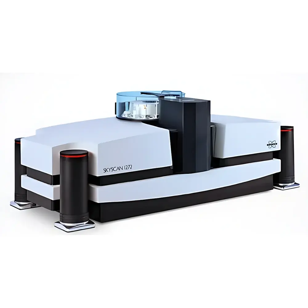
The Bruker SkyScan 1272 is a high-resolution, benchtop X-ray micro-computed tomography (micro-CT) system engineered for non-destructive, three-dimensional structural analysis of biological specimens—including intact plants, small animals, excised organs, and biomaterials—under controlled environmental or mechanical conditions. Operating on the principle of cone-beam X-ray tomography, the system acquires hundreds of projection images as the sample rotates 360°, followed by iterative or filtered-backprojection reconstruction to generate isotropic volumetric datasets with sub-micron spatial resolution. Its core architecture integrates a sealed, maintenance-free microfocus X-ray source, a large-format 16-megapixel scientific CMOS (sCMOS) detector, and precision motorized stages—all housed in a compact, self-shielded enclosure requiring only a standard 110–240 V AC outlet and no external cooling or compressed air. Designed for routine use in academic, pharmaceutical, and agricultural research laboratories, the SkyScan 1272 delivers clinical-grade image fidelity at preclinical scale, enabling quantitative morphometric assessment without histological sectioning or staining.
Key Features
- Ultra-High Resolution Imaging: Achieves isotropic voxel sizes down to 0.4 µm using geometric magnification and a high-DQE sCMOS detector with 11,200 × 11,200 native pixel resolution—enabling visualization of plant vasculature, root cortical aerenchyma, seed endosperm microstructure, and murine alveolar septa.
- Clean Image™ Acquisition Mode: Reduces beam-hardening, ring, and motion artifacts at acquisition level via optimized source-filter-detector geometry and real-time background correction—minimizing reliance on post-reconstruction software correction.
- Genius Mode Automation: Automatically selects optimal parameters—including tube voltage, current, filter selection, exposure time, and magnification—based on sample density and size; maximizes signal-to-noise ratio by dynamically positioning the sample close to the focal spot.
- Benchtop In Situ Capability: Integrated mechanical testing stages support uniaxial compression (up to 4400 N) and tension (up to 440 N); thermal stages enable temperature-controlled scanning from −30 °C to +80 °C—all recognized automatically by the control software without external cabling.
- High-Throughput Workflow: Optional 16-position automated sample changer enables unattended batch scanning with LED status feedback and auto-detection of loaded samples—critical for longitudinal plant phenotyping or genetic screening cohorts.
Sample Compatibility & Compliance
The SkyScan 1272 accommodates a broad range of biological specimens without destructive preparation: intact Arabidopsis rosettes, maize roots, rice panicles, zebrafish embryos, murine lungs, and ex vivo bone segments. Sample diameters up to 25 mm are supported within the field of view. For soft-tissue contrast enhancement, iodine-based contrast agents (e.g., Lugol’s solution) or critical-point drying protocols may be applied prior to scanning. The system complies with IEC 61000-6-3 (EMC) and IEC 61000-6-4 (emission) standards; its radiation shielding meets EU Directive 2013/59/Euratom requirements for Class IIb X-ray equipment. Data integrity and audit trail functionality in 3D.SUITE align with GLP and FDA 21 CFR Part 11 principles when configured with user authentication and electronic signature modules.
Software & Data Management
The bundled 3D.SUITE software platform provides a fully integrated workflow—from raw projection reconstruction to quantitative morphometry. NRecon performs GPU-accelerated filtered back-projection and iterative reconstruction (SIRT, SART). CTAnalyser implements standardized ASBMR-compliant bone morphometry (BV/TV, Tb.Th, Tb.Sp, Conn.D), lung tissue segmentation (using adaptive thresholding and region-growing), and plant trait extraction (root length density, xylem vessel diameter distribution, leaf vein network topology). Phase-contrast retrieval (Paganin method) enhances edge contrast in low-absorption specimens such as hydrated leaves or embryonic tissues. All processing steps are scriptable via Python API, and metadata—including acquisition parameters, stage positions, and calibration references—is embedded in DICOM and TIFF-based output formats for FAIR data management.
Applications
- Plant Phenomics: Time-resolved 3D imaging of root architecture dynamics under drought or nutrient stress; quantification of aeration pore networks in flood-tolerant rice varieties; vascular bundle patterning in developing stems.
- Preclinical Bone Research: Longitudinal monitoring of trabecular degradation in osteoporosis models; assessment of cortical porosity following glucocorticoid treatment; evaluation of scaffold integration in bone regeneration studies.
- Pulmonary Structural Analysis: Quantitative emphysema index calculation via local thickness mapping; airway wall thickening metrics in asthma models; tumor volume and necrosis fraction estimation in orthotopic lung cancer xenografts.
- Developmental Biology: Embryonic organogenesis in zebrafish and Xenopus; neural tube closure defects; cardiac chamber morphology in mouse mutants—without sectioning artifacts or antibody penetration limitations.
- Seed & Tissue Quality Control: Detection of internal voids, insect infestation, or fungal colonization in cereal grains; assessment of freeze-thaw damage in cryopreserved germplasm.
FAQ
What is the minimum achievable voxel size, and what factors influence it?
The practical voxel size depends on geometric magnification, source focal spot size, and detector pixel pitch. Under optimal setup (maximum magnification, high-resolution mode), the system achieves isotropic voxels down to 0.4 µm. Actual resolution is verified using ISO 15739-based modulation transfer function (MTF) measurements with calibrated line-pair phantoms.
Can the system perform longitudinal in vivo scans on live plants or small animals?
Yes—when used with low-dose protocols and appropriate immobilization (e.g., custom plant holders or anesthesia-compatible animal beds), the SkyScan 1272 supports repeated scanning over days or weeks. Dose optimization tools in NRecon help balance image quality against cumulative radiation exposure.
Is phase-contrast imaging supported natively?
Yes—the Paganin algorithm is implemented in CTAnalyser for propagation-based phase retrieval, significantly improving soft-tissue contrast in low-absorption samples such as hydrated leaves, young seedlings, or unstained brain tissue.
How is calibration maintained across long-term deployments?
The system includes built-in reference phantoms for attenuation calibration (hydroxyapatite rods for BMD scaling) and spatial calibration (precision ball grids). Daily warm-up routines and quarterly verification protocols ensure stability per ISO/IEC 17025-aligned laboratory quality systems.
Does the software support batch processing for large-scale phenotyping studies?
Yes—CTAnalyser supports macro scripting and command-line execution. Combined with the 16-position auto-sampler, users routinely process >100 plant or animal samples per week with consistent parameter sets and automated report generation in CSV and PDF formats.


