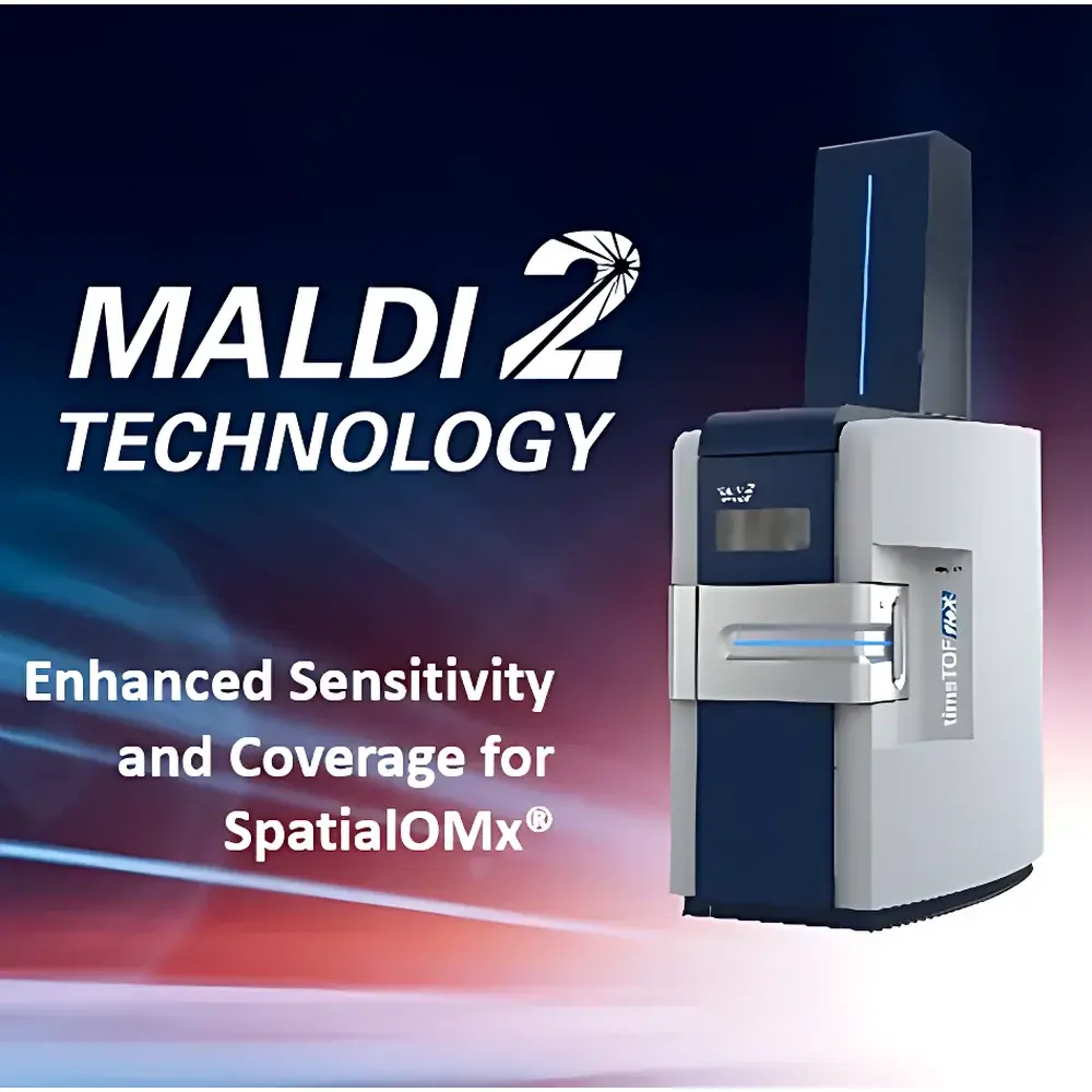
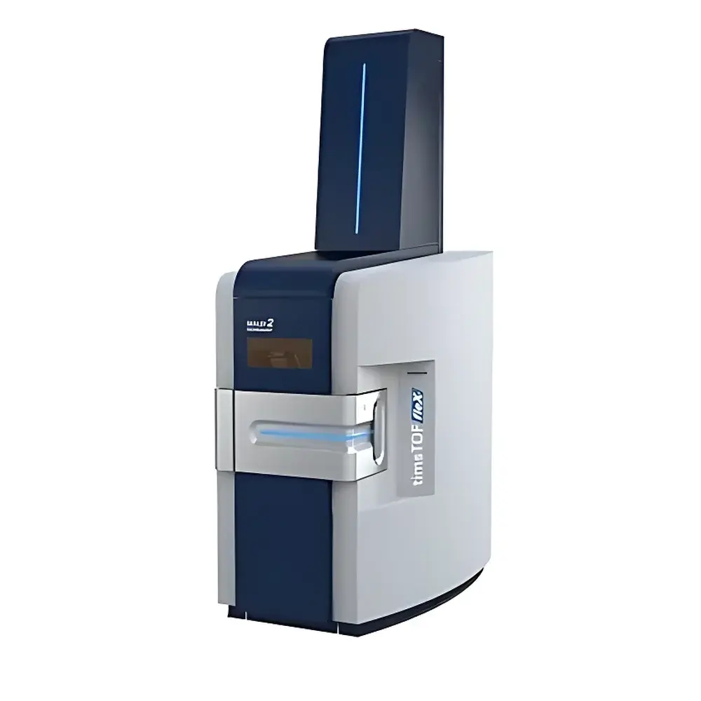
Bruker timsTOF fleX with MALDI-2
| Brand | Bruker |
|---|---|
| Origin | Germany |
| Instrument Type | Ion Mobility Mass Spectrometer |
| Model | timsTOF fleX with MALDI-2 |
| Application Scope | Universal (Biological Tissue Imaging, Lipidomics, Metabolomics, Drug Distribution Studies) |
| Regulatory Compliance | Designed for GLP/GMP-aligned workflows |
| Software Platform | SCiLS Lab, DataAnalysis, and TIMS Control integrated in Compass 6.0 suite |
| Ionization Modes | MALDI and MALDI-2 (laser post-ionization), ESI (via LC coupling) |
Overview
The Bruker timsTOF fleX with MALDI-2 is a high-performance hybrid ion mobility–time-of-flight mass spectrometer engineered for deep molecular phenotyping of biological tissues and complex samples. At its core, the system integrates trapped ion mobility spectrometry (TIMS) with orthogonal time-of-flight (TOF) detection, enabling simultaneous separation by collision cross-section (CCS), m/z, and retention time (when coupled to LC). The optional MALDI-2 module introduces a pulsed laser-based post-ionization step following conventional MALDI desorption—effectively converting neutral desorbed species into gas-phase ions. This dual-stage ionization mechanism overcomes fundamental limitations of standard MALDI, particularly low ionization efficiency and severe suppression effects in heterogeneous tissue matrices. As a result, the platform delivers enhanced sensitivity (up to 100× improvement over conventional MALDI), expanded analyte coverage—including lipids, metabolites, peptides, and small-molecule drugs—and improved quantitative reproducibility across spatially resolved imaging experiments.
Key Features
- Integrated TIMS-TOF architecture providing CCS as a fourth analytical dimension alongside m/z, retention time, and spatial coordinates
- MALDI-2 capability: non-invasive, software-switchable laser post-ionization that operates without hardware modification or recalibration
- High-speed, high-resolution imaging: pixel acquisition rates up to 100 Hz with sub-50 µm spatial resolution (dependent on laser focus and matrix application)
- Dual-source flexibility: seamless transition between MALDI, MALDI-2, and electrospray ionization (ESI) for complementary LC-MS/MS validation
- Compass 6.0 software environment with embedded TIMS Control, SCiLS Lab for multivariate image analysis, and DataAnalysis for spectral processing and CCS calibration
- Full compliance with ISO/IEC 17025 principles for method validation; supports audit-trail-enabled workflows per FDA 21 CFR Part 11 when configured with bruker.bss server infrastructure
Sample Compatibility & Compliance
The timsTOF fleX with MALDI-2 is validated for use with formalin-fixed paraffin-embedded (FFPE) and fresh-frozen tissue sections (typically 5–20 µm thickness), single-cell suspensions, microbial colonies, and plant tissue slices. Compatible substrates include conductive ITO-coated slides, stainless steel IntelliSlides, and functionalized polymer surfaces. All MALDI matrix formulations supported by Bruker—including α-cyano-4-hydroxycinnamic acid (CHCA), 2,5-dihydroxybenzoic acid (DHB), and 9-aminoacridine (9-AA)—are fully operational under MALDI-2 conditions. The system meets essential requirements for regulated bioanalysis: it supports documented instrument qualification (IQ/OQ/PQ), method transfer protocols aligned with USP , and long-term stability testing per ICH Q2(R2). Routine operation adheres to GLP principles for preclinical imaging studies and GMP-aligned biomarker discovery pipelines.
Software & Data Management
Data acquisition and interpretation are unified within the Compass 6.0 software ecosystem. TIMS Control manages mobility separation parameters (e.g., ramp time, accumulation time, elution voltage), while SCiLS Lab enables unsupervised segmentation, statistical mapping (e.g., PCA, t-SNE), and co-localization analysis across multi-omic datasets. All raw data (.d format) are stored with embedded metadata including laser fluence, detector gain, mobility calibration standards, and user-defined annotation layers. Export options include imzML for third-party tools (e.g., MSiReader, Cardinal), mzML for ProteoSAFe integration, and HDF5 for scalable cloud-based processing. Audit trails record every parameter change, file export, and reprocessing event—ensuring full traceability required for regulatory submissions.
Applications
- Spatially resolved lipidomics and metabolomics in brain, liver, and tumor tissues
- Drug and metabolite distribution mapping across dose-response gradients in pharmacokinetic/pharmacodynamic (PK/PD) studies
- Endogenous peptide imaging for neurodegenerative disease biomarker discovery
- Microbiome–host interface analysis via direct tissue surface profiling
- Validation of LC-MS/MS findings through orthogonal spatial correlation using CCS-filtered ion images
- Multi-modal integration with histopathology (H&E staining) and digital pathology platforms
FAQ
How does MALDI-2 differ from conventional MALDI in terms of ionization mechanism?
MALDI-2 employs a secondary UV laser pulse delivered nanoseconds after the primary MALDI desorption pulse, ionizing neutrals released during ablation—thereby increasing total ion yield and reducing matrix-related suppression.
Is hardware modification required to enable MALDI-2 on an existing timsTOF fleX system?
No. MALDI-2 is activated entirely through software configuration and requires only firmware update and calibration with proprietary reference standards.
What types of analytes benefit most from MALDI-2 enhancement?
Compounds with low proton affinity or high polarity—such as phospholipids, glycerolipids, organic acids, and phase II drug conjugates—show the greatest signal enhancement (2–3 orders of magnitude).
Can CCS values obtained with MALDI-2 be directly compared to those from LC-MALDI or nanoESI experiments?
Yes. CCS is an intrinsic physicochemical property independent of ionization mode; TIMS-derived CCS values are fully transferable across platforms when calibrated using the same polyalanine or tune mix standards.
Does the system support automated quantification in imaging mode?
Quantitative imaging is enabled via internal standard normalization (e.g., deuterated lipid spikes) and response factor correction derived from parallel LC-MS/MS calibration curves—fully implemented in SCiLS Lab’s Quant Module.
Related Products

")