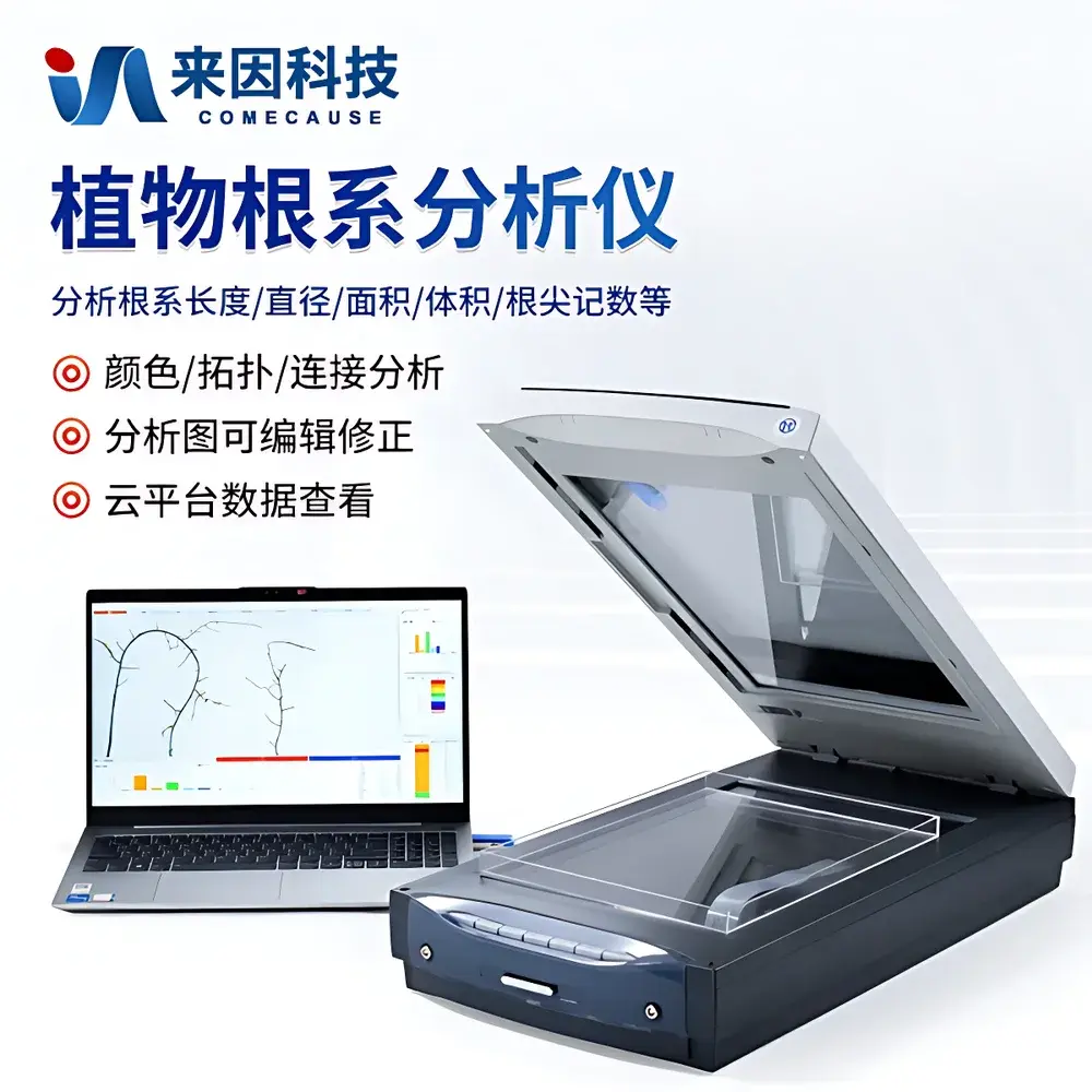
COMECAUSE IN&GX02 High-Resolution Dual-Illumination Root Imaging & Morphometric Analysis System
| Brand | COMECAUSE |
|---|---|
| Origin | Shandong, China |
| Manufacturer | Yes |
| Type | Domestic |
| Model | IN&GX02 |
| Price | USD 2,950 (FOB) |
| Optical Resolution | 4800 × 9600 dpi (A4+ format) |
| Scan Area (Reflective) | 355.6 mm × 215.9 mm |
| Scan Area (Transmissive) | 320.0 mm × 203.2 mm |
| Minimum Pixel Size | 0.005 mm × 0.0026 mm |
| Image Formats Supported | TIFF, JPEG |
| Software Licensing | Hardware-encrypted USB dongle |
| Operating System | Windows 10 Pro/Enterprise (64-bit) |
| Interface | USB 2.0 |
| Light Source | Dual cold-cathode fluorescent lamp (CCFL), white spectrum |
| Bit Depth | 48-bit color |
| Topological Analysis | Root hierarchy mapping, branching angle, connection count, root-order extraction |
| Fractal Analysis | Box-counting dimension calculation |
| Color-Based Segmentation | Multi-threshold RGB/HSL segmentation for functional root classification |
| Output Export | Excel (.xlsx), annotated image overlays, distribution histograms, cloud-synced project archives |
Overview
The COMECAUSE IN&GX02 High-Resolution Dual-Illumination Root Imaging & Morphometric Analysis System is a laboratory-grade instrumentation platform engineered for non-destructive, high-fidelity digital phenotyping of excised plant root systems. It operates on the principle of contact-based optical imaging—leveraging precisely calibrated dual-side illumination and sub-5-µm spatial resolution to capture distortion-free, shadow-minimized grayscale or color reflectance/transmission scans of washed root samples or rhizobox-grown specimens. Unlike destructive excavation or low-resolution imaging methods, this system enables quantitative morphometric reconstruction from static 2D projections, supporting rigorous analysis of root architecture under controlled experimental conditions. Its design aligns with established protocols in plant phenomics, root ecology, and crop physiology research where reproducible, operator-independent measurement of structural traits is essential for genotype–phenotype correlation studies, drought-response screening, nutrient-use efficiency evaluation, and symbiotic interaction quantification (e.g., rhizobia nodule volume fraction).
Key Features
- Dual-illumination A4+ scanning module (355.6 × 215.9 mm reflective area) with independently controlled CCFL light sources above and below the transparent root tray—eliminating parallax, cast shadows, and uneven contrast typical in single-source setups.
- Optical resolution of 4800 × 9600 dpi (0.005 mm × 0.0026 mm pixel pitch), enabling reliable detection and segmentation of fine lateral roots ≥ 50 µm in diameter.
- Hardware-encrypted software suite with automated root skeletonization, topology graph generation, and hierarchical root-order assignment—supporting both whole-system and branch-specific parameter extraction.
- Comprehensive morphometric engine delivering 22 standardized output metrics including total root length, average/diameter median/max, surface area, volume (via cylinder approximation), tip count, branching frequency, overlap correction, fractal dimension (box-counting method), and angular distribution (gravitropic/horizontal orientation angles).
- Multi-channel color analysis module permitting spectral segmentation of root segments by hue, saturation, and luminance—enabling differentiation of metabolically active zones, senescing tissues, mycorrhizal colonization regions, and nitrogen-fixing nodules.
- Batch-processing capability with editable annotation layers, manual correction tools, and project-level metadata tagging compliant with MIAPPE (Minimum Information About a Plant Phenotyping Experiment) guidelines.
Sample Compatibility & Compliance
The IN&GX02 system accepts root samples prepared via standard washing protocols (e.g., soil suspension sieving, hydroponic harvesting) or grown in transparent rhizoboxes (acrylic or glass). Compatible specimen types include monocots (e.g., rice, maize), dicots (e.g., Arabidopsis, soybean, tomato), and perennial woody species when trimmed to fit the scan area. All analyses adhere to ISO 21527-1:2008 (microbiological methods for roots) and ASTM D7789-15 (standard guide for root architecture characterization) for measurement traceability. Data audit trails—including timestamped scan logs, user ID, calibration records, and software version—meet GLP-compliant documentation requirements. Optional cloud synchronization supports remote collaboration and long-term data integrity per FAIR principles (Findable, Accessible, Interoperable, Reusable).
Software & Data Management
The proprietary analysis software runs natively on Windows 10 (64-bit) and requires no internet connectivity for core computation. It employs deterministic, non-statistical image processing algorithms—avoiding stochastic thresholding—to ensure inter-laboratory reproducibility. Each analysis session generates a structured project archive containing raw TIFF/JPEG files, binary mask layers, skeleton graphs, topological adjacency matrices, and tabular outputs (.xlsx). All results are exportable with embedded units (mm, mm², mm³, °) and SI-compliant prefixes. Cloud integration (optional subscription) provides encrypted AES-256 storage, role-based access control, and versioned backups synchronized across devices. The interface supports bilingual operation (English/Chinese) with one-click language switching—no reinstallation required.
Applications
- Quantitative root phenotyping in forward/reverse genetic screens, QTL mapping, and genome-wide association studies (GWAS).
- Evaluation of abiotic stress responses—including water deficit, salinity, aluminum toxicity, and phosphorus limitation—via changes in root depth distribution, lateral density, and diameter-class allocation.
- Assessment of symbiotic efficacy: volumetric quantification of rhizobial nodules relative to host root biomass; spatial mapping of arbuscular mycorrhizal hyphal penetration zones.
- Validation of root growth models (e.g., CRootBox, SimRoot) using empirically derived architectural parameters as boundary conditions.
- Curriculum-integrated teaching modules in plant science laboratories, emphasizing digital literacy, image-based reasoning, and open-data practices.
- Regulatory submission support for biopesticide or biostimulant trials requiring root morphology endpoints per OECD TG 208 or EPPO PP1/263(1) guidelines.
FAQ
What sample preparation is required prior to scanning?
Root systems must be thoroughly cleaned of soil particles using gentle agitation in deionized water and blotted dry. For rhizobox-grown specimens, roots should remain intact on the transparent barrier; no excision is needed.
Can the system analyze live roots in situ?
No. The IN&GX02 is designed for ex situ analysis of excised or rhizobox-confined roots. It does not support real-time in-soil imaging; for dynamic monitoring, complementary techniques such as minirhizotrons or X-ray CT are recommended.
Is FDA 21 CFR Part 11 compliance supported?
The standalone software does not include electronic signature or audit trail modules required for Part 11. However, full audit log exports (CSV/Excel) and immutable project archives enable manual implementation within validated institutional workflows.
What computing resources are mandatory?
Minimum: Intel Core i5-9400 / 16 GB RAM / 256 GB SSD / Windows 10 Pro 64-bit / 4× USB 2.0 ports. Recommended: i7-10700K / 32 GB RAM / dedicated GPU for accelerated batch processing.
Are calibration standards provided with the system?
Yes—a certified NIST-traceable resolution target and grayscale step wedge are included for routine optical verification and intensity normalization before each analytical session.


