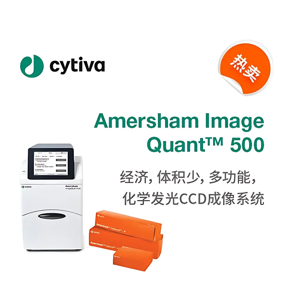
Cytiva Amersham ImageQuant™ 500 Chemiluminescent CCD Gel Imaging System
| Brand | Cytiva |
|---|---|
| Origin | Japan |
| Model | Amersham ImageQuant 500 |
| Imaging Type | Chemiluminescence |
| CCD Resolution | 8.3 MP |
| Dynamic Range | 4.8 decades |
| Lens Aperture | f/0.14 (Fujifilm) |
Overview
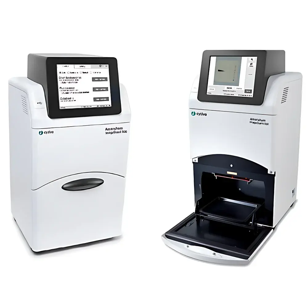
The Cytiva Amersham ImageQuant™ 500 Chemiluminescent CCD Gel Imaging System is a purpose-engineered, compact benchtop imager designed for quantitative detection and documentation of chemiluminescent, fluorescent, and visible-light signals in life science applications. Built around a thermoelectrically cooled 8.3-megapixel CCD sensor with Fujifilm optical components—including an ultra-fast f/0.14 lens—the system delivers high-sensitivity imaging across multiple modalities: Western blot chemiluminescence (down to picogram-level detection), SYBR™ Green- or ethidium bromide-stained nucleic acids under UV/blue light excitation, total protein staining (e.g., Deep Purple), and colorimetric assays under white light. Its integrated Peltier cooling architecture achieves stable sensor temperatures ≤ –25 °C within five minutes of power-on, enabling low-noise acquisition essential for high dynamic range (4.8 decades) and linear signal quantification—critical for comparative densitometry and regulatory-compliant workflows.
Key Features
- Thermoelectrically cooled 8.3 MP CCD sensor with real-time noise suppression via pixel binning and dark-current compensation
- Fujifilm f/0.14 fixed-focus lens optimized for maximum photon capture in low-light chemiluminescent applications
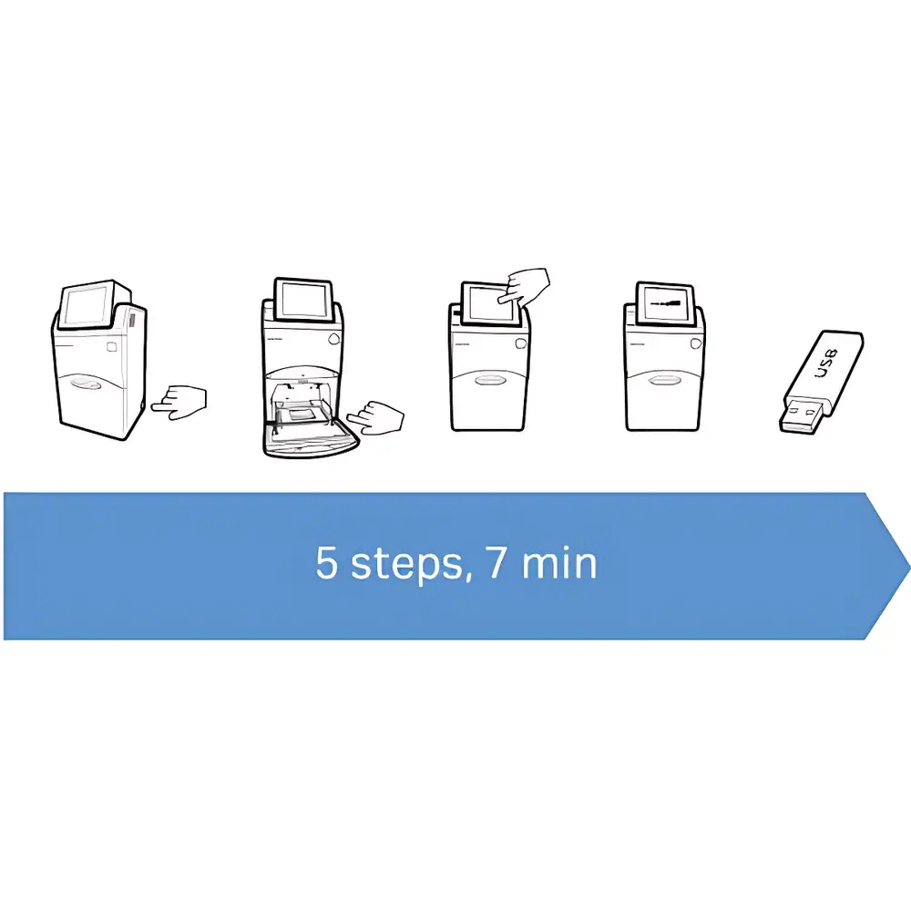
- Integrated 10.1-inch capacitive touchscreen interface—enables fully standalone operation without external PC or dedicated software installation
- Triple LED illumination module: side-mounted UV (302 nm), blue light (470 nm), and white light sources—individually controllable for multiplexed fluorescence and transillumination
- Auto-color overlay functionality: simultaneous acquisition and precise spatial registration of chemiluminescent blots and colorimetric molecular weight markers for accurate band annotation
- Flexible exposure control: manual single-shot, auto-exposure, semi-automatic region-of-interest (ROI)-guided exposure, and multi-frame accumulation mode
- Onboard image storage to internal SSD, USB flash drives, or network-mounted SMB/CIFS shares—supports DICOM export and TIFF/PNG/JPEG formats with embedded metadata
Sample Compatibility & Compliance
The ImageQuant 500 accommodates standard electrophoresis formats including mini-gels (up to 10 × 10 cm), large-format Western blots (up to 15 × 20 cm), and membrane-based immunoassays. It supports all major chemiluminescent substrates (e.g., ECL, SuperSignal, Luminata), DNA intercalating dyes (ethidium bromide, SYBR Safe, SYBR Gold), total-protein stains (Deep Purple, Coomassie Blue), and chromogenic substrates (BCIP/NBT, DAB). The system’s hardware design and firmware architecture comply with GLP/GMP-aligned data integrity principles: audit-trail-enabled user authentication, timestamped acquisition logs, and immutable image metadata (exposure time, gain, lens aperture, illumination source, temperature) are embedded directly into exported files. While not certified as FDA 21 CFR Part 11 compliant out-of-the-box, the system is fully compatible with validated ImageQuant TL software deployments that support electronic signatures, role-based access control, and full audit trail generation for regulated environments.
Software & Data Management
The imager operates natively via its embedded Linux-based firmware and touchscreen UI—no host PC required for acquisition. For advanced analysis, it integrates seamlessly with Cytiva’s ImageQuant TL v8.4+ software (sold separately), which provides lane/band detection, background subtraction, molecular weight calibration, relative quantification, and statistical reporting. All acquired images retain EXIF-like metadata fields—including exposure parameters, sensor temperature, and illumination configuration—ensuring traceability and reproducibility. Data export options include lossless 16-bit TIFF (with LZW compression), PNG, JPEG, and DICOM format for PACS integration. Network connectivity supports LDAP authentication, SMB file sharing, and automated backup to centralized NAS or LIMS repositories.
Applications
- Quantitative Western blotting: detection and densitometric analysis of chemiluminescent signals from HRP- or AP-conjugated secondary antibodies
- Nucleic acid gel documentation: high-sensitivity imaging of ethidium bromide-, SYBR™ Green-, or GelRed-stained DNA/RNA gels under UV or blue light
- Total protein normalization: uniform visualization of loading controls using membrane-compatible total protein stains
- Multiplexed blot analysis: co-registration of chemiluminescent target bands with pre-stained colorimetric ladders for unambiguous MW assignment
- Cell-based assay documentation: imaging of colorimetric ELISA plates, colony formation assays, or viability stains under white light
- Teaching and core facility use: intuitive workflow reduces training time; compact footprint maximizes bench space efficiency
FAQ
What is the minimum detectable amount of protein using chemiluminescence on this system?
Typical detection limits range from 1–5 pg of horseradish peroxidase (HRP)-conjugated antibody bound to target antigen under optimized substrate conditions—dependent on antibody affinity, substrate kinetics, and membrane type.
Does the system support time-lapse or kinetic chemiluminescence imaging?
No—it is optimized for endpoint static imaging. Kinetic acquisition requires external PC-based control and is not supported by the onboard firmware.
Can I perform background subtraction or lane normalization directly on the instrument?
Basic ROI-based intensity measurement and manual background correction are available on the touchscreen interface; full densitometry, lane normalization, and statistical analysis require ImageQuant TL software.
Is the UV light source suitable for direct DNA crosslinking?
No—the 302 nm UV LED is calibrated solely for transillumination and visualization; it lacks the irradiance and spectral output required for effective UV crosslinking.
How is calibration maintained across instruments and over time?
The system includes factory-calibrated reference standards for intensity linearity and spatial uniformity; users may perform optional daily flat-field correction using the built-in white light source to compensate for optical drift.
Related Products


