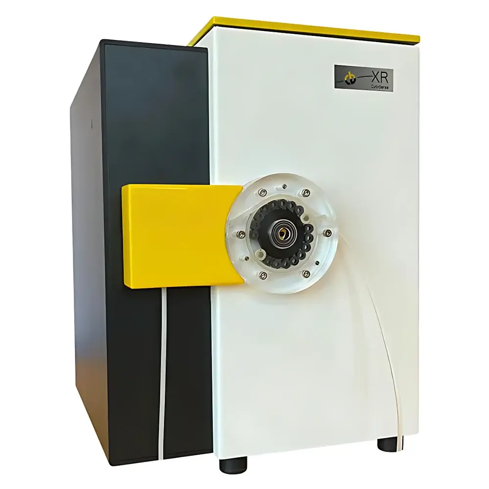
CytoBuoy CytoSense XR Scanning Imaging Flow Cytometer for Phytoplankton Community Analysis
| Brand | CytoBuoy (Netherlands) |
|---|---|
| Origin | Netherlands |
| Model | CytoSense XR |
| Detection Principle | Laser-scanning flow cytometry with hydrodynamic focusing and synchronized high-resolution imaging |
| Particle Size Range | 0.2–700 µm |
| Concentration Range | 10²–10¹¹ particles/L |
| Sheath Fluid System | Closed-loop recirculating |
| Optical Channels | Forward scatter (FWS), red fluorescence (FLRed, ~680 nm), side scatter (optional), multi-wavelength excitation (488 nm standard, optional 532/640 nm) |
| Imaging Mode | Pulse-profile scanning + triggered micrograph acquisition |
| Data Output | Per-particle parameter vectors (size, shape, pigment distribution, internal structure morphology), time-stamped event files, TIFF/JPEG image stacks |
| Compliance | Designed for GLP-compliant environmental monitoring |
Overview
The CytoBuoy CytoSense XR is a field-deployable, scanning imaging flow cytometer engineered specifically for quantitative phytoplankton community analysis in natural aquatic systems. Unlike clinical-grade flow cytometers—designed for narrow-diameter tubing and homogeneous mammalian cells—the CytoSense XR employs a wide-bore, low-shear fluidic architecture optimized for heterogeneous, fragile, and aggregated particles found in freshwater, estuarine, and marine environments. Its core measurement principle combines hydrodynamic focusing with pulsed laser scanning (488 nm excitation) and synchronized optical profiling. As particles traverse the measurement zone at laminar flow velocities, they are interrogated by a focused laser beam orthogonal to the flow axis. Each particle generates forward scatter (FWS), side scatter (SSC), and chlorophyll-specific red fluorescence (FLRed), while simultaneously producing a one-dimensional optical “pulse profile” that encodes axial dimension, internal pigment distribution (e.g., chloroplast positioning), and morphological features such as chain integrity in diatoms or filament continuity in cyanobacteria. This pulse-based signature—distinct from conventional gated intensity measurements—enables robust classification independent of particle orientation or rotational drift, a critical advantage for unfiltered, in-situ water samples.
Key Features
- Full-size-range analysis without pre-filtration: Direct quantification of particles from 0.2 µm (bacteria) to 700 µm (colonial algae, copepod nauplii, detrital aggregates)
- Low-shear fluidics: Capable of intact analysis of delicate structures—including Microcystis colonies, Phaeocystis blooms, and filamentous cyanobacteria up to 2.5 mm in length
- Pulse-profile scanning: Captures amplitude, duration, and shape of optical signals along the particle’s travel axis—providing subcellular resolution of pigment localization and structural heterogeneity
- Integrated imaging module: Triggered micrograph acquisition synchronized with optical pulses; supports both “smart grid” (fluorescence/scatter-triggered) and user-defined subset targeting modes
- Closed-loop sheath fluid system: Minimizes contamination risk, eliminates external waste discharge, and ensures stable hydraulic performance across extended deployments
- Automated cleaning protocol: Integrated rinse cycles reduce maintenance intervals and support unattended operation in remote or buoy-mounted configurations
Sample Compatibility & Compliance
The CytoSense XR accepts raw, unprocessed water samples—including turbid reservoirs, eutrophic lakes, coastal seawater, and wastewater effluents—without centrifugation, filtration, or dilution. It reliably resolves taxonomically relevant features across prokaryotes (Synechococcus, Prochlorococcus), eukaryotic phytoplankton (diatoms, dinoflagellates, cryptophytes), colonial forms (Anabaena, Gonyaulax), microzooplankton (ciliates, rotifers), and non-biological particulates (TEP, microplastics, mineral sediments). The instrument meets functional requirements for environmental monitoring under ISO 14442 (water quality—phytoplankton enumeration), ASTM D5905 (microbiological analysis of water), and aligns with data integrity expectations outlined in FDA 21 CFR Part 11 when deployed with validated software configurations. Its closed fluid path and traceable calibration protocols support GLP-compliant reporting frameworks used by national water authorities and research consortia.
Software & Data Management
CytoSense software (v6.x+) provides a unified environment for real-time acquisition, pulse-profile visualization, scatter/fluorescence gating, and supervised clustering using hierarchical agglomerative algorithms. Each detected particle is stored with full parameter vectors—including FWS peak height, FLRed integral, pulse width, skewness, and image metadata—and exported in standardized HDF5 or CSV formats. The software supports library-based classification: users may curate reference image libraries from known monocultures or field isolates, then apply machine-learning-assisted matching to new samples for automated genus- or species-level annotation. Audit trails log all processing steps, parameter adjustments, and operator actions. Exported datasets are compatible with R (cytofR), Python (FlowKit), and MATLAB toolchains for advanced multivariate analysis, time-series trend modeling, and early-warning algorithm development.
Applications
- Real-time phytoplankton community monitoring in drinking water intakes, reservoirs, and aquaculture facilities
- Detection and quantification of harmful algal bloom (HAB) taxa—including Alexandrium, Pseudo-nitzschia, and microcystin-producing Microcystis—prior to toxin accumulation
- Long-term ecological assessment of climate-driven shifts in plankton composition and size spectra
- Validation of satellite-derived chlorophyll-a estimates through in-situ particle-specific pigment quantification
- Research on trophic interactions, including grazing pressure assessments via gut-content imaging of microzooplankton
- Microplastic and TEP quantification in biogeochemical cycling studies
FAQ
Can the CytoSense XR operate autonomously in remote field locations?
Yes. With optional power management modules (solar/battery), telemetry integration (4G/LoRaWAN), and scheduled sampling protocols, it supports unattended deployment for weeks to months.
Does it require daily calibration or daily maintenance?
No. The closed-loop sheath system and automated cleaning routines enable stable operation for >72 hours between manual interventions; annual optical alignment verification is recommended.
How does it differentiate morphologically similar species, such as Thalassiosira versus Coscinodiscus?
Through combined analysis of pulse-profile asymmetry, FWS/FLRed ratio gradients, and internal pigment banding patterns—features resolvable only via axial scanning, not conventional flow cytometry.
Is raw image data export supported for third-party AI training?
Yes. Full-resolution TIFF stacks and associated parameter metadata are exportable without proprietary compression or obfuscation.
Can it be integrated into existing SCADA or LIMS platforms?
Yes. Native support for OPC UA, MQTT, and RESTful API enables bidirectional data exchange with industrial control systems and laboratory information management systems.


