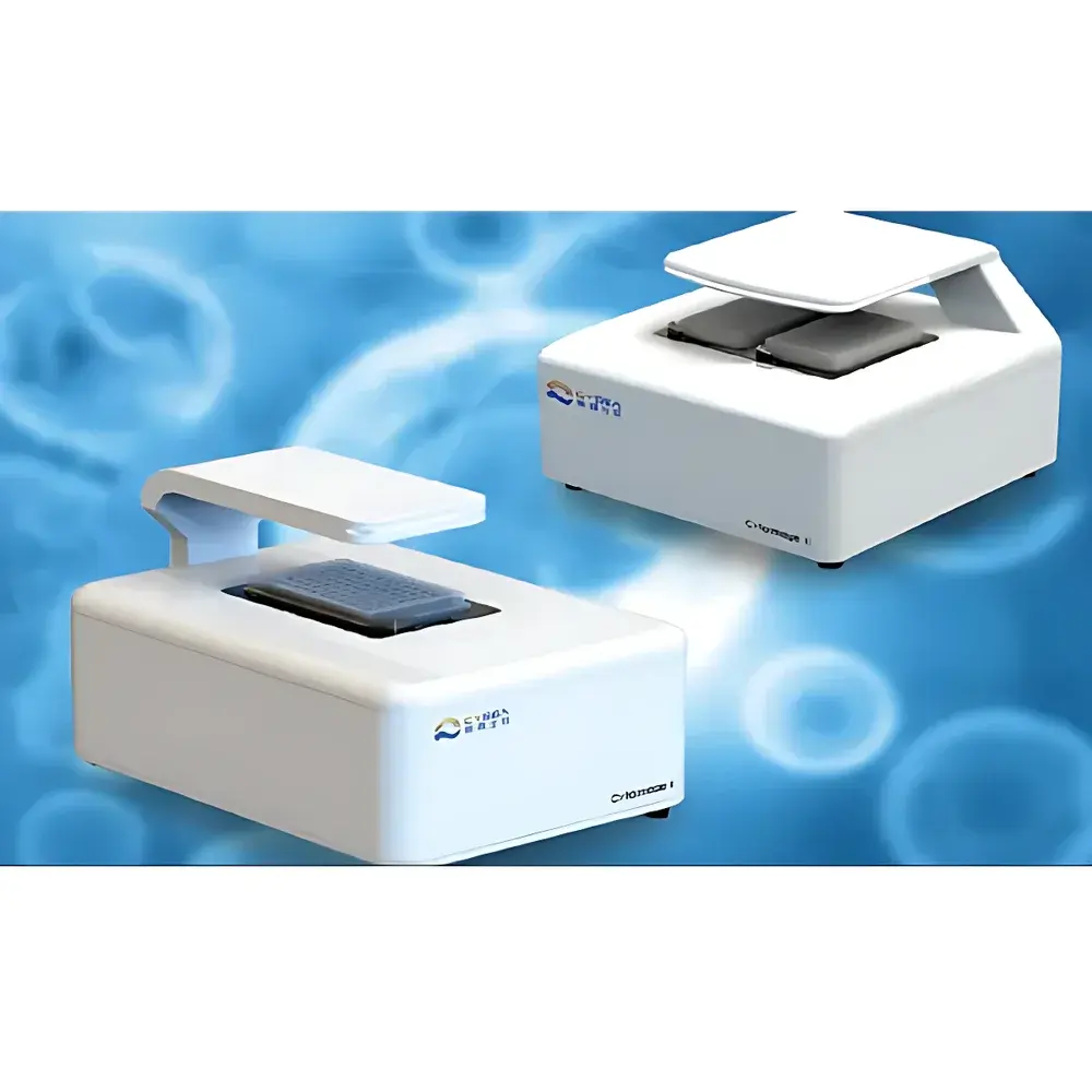
Cytoimage I Live-Cell Imaging System by CYSEA
| Brand | CYSEA |
|---|---|
| Origin | Beijing, China |
| Manufacturer Type | Original Equipment Manufacturer (OEM) |
| Product Origin | Domestic (China) |
| Model | Cytoimage I |
| Pricing | Upon Request |
Overview
The Cytoimage I Live-Cell Imaging System is an integrated, incubator-compatible, fully automated microscopy platform engineered for long-term, non-invasive, quantitative observation of living cells under physiologically relevant conditions. Unlike conventional benchtop microscopes requiring repeated removal of samples from controlled environments, the Cytoimage I is designed to operate continuously inside standard CO₂ incubators—withstanding ambient temperatures up to 37 °C, relative humidity ≥95%, and mildly acidic atmospheres (pH 6.8–7.4) without optical or mechanical degradation. Its core architecture employs a fixed-stage, lens-scanning optical design: instead of moving delicate cell cultures on motorized stages, high-precision piezoelectric actuators translate the objective lens assembly across the sample plane. This eliminates mechanical perturbation, thermal drift, and positional uncertainty associated with stage-based systems—critical for maintaining cellular integrity during time-lapse experiments spanning hours to days.
Key Features
- Incubator-integrated operation: IP54-rated enclosure with corrosion-resistant housing and sealed optical pathways for reliable performance in humid, CO₂-rich environments.
- Lens-scanning autofocus and positioning: Eliminates sample displacement; enables sub-micron repeatability (<0.3 µm RMS positional error) across hundreds of acquisition cycles.
- Multi-site, multi-well dynamic imaging: Supports automated acquisition from up to 384 wells (e.g., 384-well plates), with independent focus and exposure control per site.
- Multi-channel fluorescence capability: Simultaneous brightfield plus up to three fluorescence channels (excitation/emission optimized for DAPI/Hoechst, FITC/GFP, and TRITC/mCherry).
- AI-assisted cell detection and tracking: On-device neural inference engine identifies morphological features (e.g., confluence, mitotic figures, neurite outgrowth) without user-defined thresholds.
- Programmable experimental workflows: Time-lapse intervals, z-stack depth, channel selection, and region-of-interest (ROI) mapping are defined via intuitive graphical protocol builder.
Sample Compatibility & Compliance
The Cytoimage I accommodates standard tissue-culture formats including 6–384-well plates, chambered coverslips, and custom microfluidic devices with ≤1.5 mm glass bottom thickness. All optical components comply with ISO 10934-1 (microscope nomenclature) and ISO 8578 (specifications for microscope objectives). The system’s software architecture supports audit trails, electronic signatures, and user-access controls aligned with GLP and GMP documentation requirements. Data export formats (TIFF, OME-TIFF, CSV) conform to NIH-supported open standards for reproducible image analysis. While not FDA-cleared as a diagnostic device, the platform meets essential requirements for preclinical research use under ISO 13485-aligned quality management systems.
Software & Data Management
The Cytoimage Control Suite provides a unified interface for instrument control, real-time preview, batch processing, and quantitative analytics. Raw image data are stored in hierarchical folder structures with embedded metadata (timestamp, environmental log, acquisition parameters). Built-in modules include confluence quantification, wound-healing assay analysis, mitosis event detection, and intensity-based kinetic profiling. All processing pipelines are scriptable via Python API (compatible with scikit-image, napari, and CellProfiler ecosystems). Audit logs record every user action—including parameter modifications, image exports, and analysis runs—with timestamps and operator IDs. Exported reports support PDF generation with configurable templates compliant with internal SOPs or external regulatory submissions.
Applications
- Longitudinal cytotoxicity screening: Real-time monitoring of membrane integrity, apoptosis markers, and proliferation kinetics across dose-response gradients.
- Stem cell differentiation assays: Tracking morphological transitions and lineage-specific marker expression over multi-day timelines.
- 3D spheroid and organoid dynamics: Z-stack-enabled growth, compaction, and necrotic core formation analysis in Matrigel-embedded cultures.
- CRISPR/Cas9 editing validation: Visual confirmation of phenotypic outcomes (e.g., loss-of-function morphology, reporter activation) without endpoint fixation.
- Microenvironmental response studies: Dual-plate comparative experiments—for example, hypoxia vs. normoxia, drug-treated vs. vehicle control—within a single incubator footprint.
FAQ
Can the Cytoimage I be used outside of a CO₂ incubator?
Yes—the system operates independently in ambient lab environments, though its thermal and humidity tolerance specifications apply only when installed inside incubators.
Does it support oil-immersion or high-NA objectives?
No—Cytoimage I is optimized for air-objective imaging (10× to 40× dry objectives); immersion optics are incompatible with its sealed, incubator-grade mechanical design.
How is focus stability maintained during extended time-lapse experiments?
Through continuous hardware-based focus feedback using infrared LED-based distance sensing and closed-loop piezo correction—no reliance on image-based autofocus algorithms.
Is remote access or cloud-based data synchronization available?
Local network access is supported via HTTPS-enabled web interface; cloud integration requires on-premise deployment of validated third-party storage solutions (e.g., NAS with SMB/NFS protocols).
What calibration standards are provided for fluorescence intensity quantification?
NIST-traceable fluorescent microsphere kits (488 nm/525 nm and 561 nm/600 nm) are included for channel-specific gain and offset calibration prior to each experimental run.


