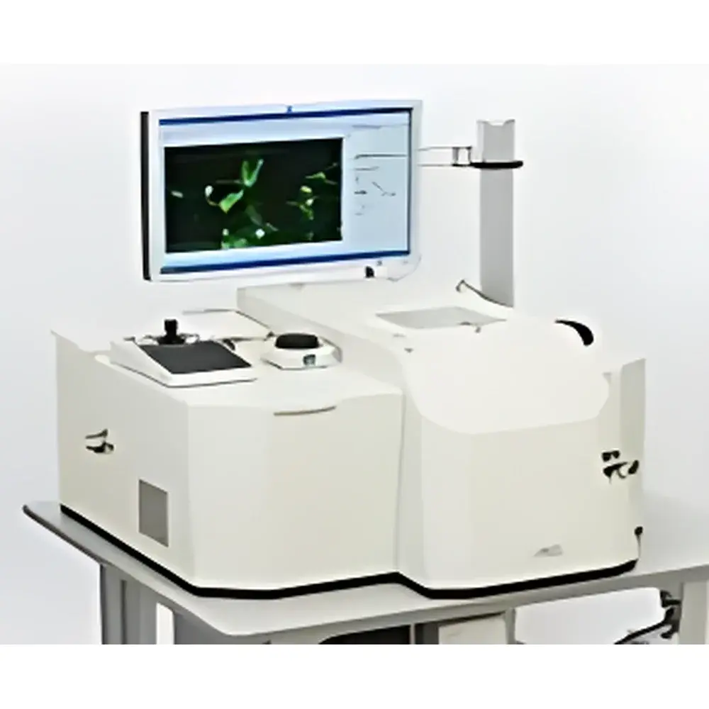
ECI TAXIScan-Fluo Fluorescent Cell Chemotaxis System
| Origin | Japan |
|---|---|
| Manufacturer Type | Authorized Distributor |
| Origin Category | Imported |
| Model | TAXIScan-Fluo |
| Pricing | Available Upon Request |
| Objective Lenses | 10×, 20×, 40×, 100× |
| Fluorescence Filter Sets | Blue (B), Green (G), Red (R) |
| Minimum Sample Requirement | ≤100 viable cells per assay |
| Temperature Control Range | Room temperature +3 °C to 40 °C |
| Microfluidic Chip Terrace Depths | 4 µm, 5 µm, 6 µm, or 8 µm |
| Parallel Assay Capacity | 12 independent microchannels |
| Autofocus | Integrated real-time autofocus system |
| Imaging Mode | Live fluorescence time-lapse microscopy with cold CCD sensor |
| Data Output Format | Movie files (e.g., AVI, TIFF stack) with metadata embedding |
| Analytical Capabilities | Automated gradient quantification, cell count, trajectory mapping, velocity vector analysis, morphometric profiling (area, perimeter, aspect ratio, circularity), directional persistence calculation |
Overview
The ECI TAXIScan-Fluo Fluorescent Cell Chemotaxis System is an advanced, microfluidics-integrated live-cell imaging platform engineered for quantitative analysis of chemotactic behavior under precisely controlled chemical gradients. Based on ECI’s proprietary silicon-based microchannel chip technology, the system enables stable, reproducible establishment of linear or nonlinear chemoattractant concentration gradients across sub-microliter volumes—without reliance on diffusion chambers or Boyden-style membranes. The core measurement principle combines microfluidic gradient generation with high-resolution epifluorescence time-lapse microscopy, permitting direct observation and statistical quantification of single-cell motility responses—including migration speed, directionality, turning frequency, pseudopod dynamics, and morphological adaptation—in real time. Designed for physiologically relevant conditions, the system maintains precise thermal regulation (±0.3 °C stability) and supports long-term imaging of primary and cultured cells without phototoxicity or environmental drift.
Key Features
- Silicon microfabricated chip with 12 parallel microchannels (each configurable with terrace depths of 4, 5, 6, or 8 µm) enabling simultaneous multi-condition assays under identical environmental parameters;
- Cold CCD imaging module positioned beneath the focal plane with coaxial epi-illumination and high-numerical-aperture optics, optimized for low-noise fluorescence detection across B/G/R spectral bands;
- Automated, software-driven autofocus maintaining sub-pixel focus stability over multi-hour acquisitions—critical for tracking subtle cytoskeletal rearrangements during polarization and migration;
- Minimal sample requirement: functional chemotaxis assays validated with as few as 50–100 viable cells per channel, reducing biological variability and conserving precious primary isolates;
- Integrated environmental control unit supporting temperature regulation from ambient +3 °C to 40 °C with active humidity compensation to prevent evaporation-induced gradient distortion;
- No darkroom required: fully shielded optical path and low-stray-light illumination architecture ensure consistent signal-to-noise ratio in standard laboratory lighting conditions.
Sample Compatibility & Compliance
The TAXIScan-Fluo accommodates a broad spectrum of adherent and non-adherent mammalian cell types, including human peripheral blood neutrophils, eosinophils, monocytes, T lymphocytes, dendritic cells, cancer cell lines (e.g., MDA-MB-231, PC-3), endothelial cells (HUVEC), neural progenitors, mesenchymal stem cells (MSCs), and induced pluripotent stem cell (iPSC)-derived models. All chip surfaces are plasma-treated and optionally coated with extracellular matrix proteins (fibronectin, collagen I/IV, laminin) to support physiological adhesion. The system complies with ISO 13485 design controls for in vitro diagnostic research instrumentation and meets essential requirements for GLP-compliant study execution. Data acquisition and analysis modules support audit trail logging, electronic signature capability, and export formats compatible with FDA 21 CFR Part 11–aligned validation protocols when deployed in regulated environments.
Software & Data Management
Acquisition and analysis are managed via ECI’s dedicated TAXIScan Analysis Suite (v5.x), a Windows-based application built on a modular, scriptable architecture. The software provides synchronized control of stage movement, illumination intensity, exposure time, z-stack acquisition, and temperature setpoints. Gradient profiles are reconstructed pixel-wise from reference dye calibration runs and embedded into each movie file’s metadata. Automated cell segmentation employs adaptive thresholding combined with machine learning–enhanced contour refinement to distinguish overlapping or densely migrating cells. Output metrics include mean migration velocity (µm/min), chemotactic index (CI = cos θ, where θ is angle between displacement vector and gradient axis), forward migration index (FMI), meandering index, and dynamic shape descriptors updated at user-defined temporal intervals (1–60 s). Raw data and processed results are exportable in CSV, HDF5, and standardized MIAME-compliant formats for integration with third-party bioinformatics pipelines.
Applications
- Chemotaxis Mechanism Studies: Quantitative dissection of GPCR- and receptor tyrosine kinase–mediated signaling cascades regulating actin polymerization, Rho GTPase activation, and front-rear polarity establishment;
- Inflammatory & Immunological Research: Functional assessment of patient-derived leukocytes in autoimmune disorders (e.g., rheumatoid arthritis, SLE) or immunodeficiency syndromes;
- Oncology & Metastasis Modeling: Evaluation of tumor cell invasion toward stromal-derived factors (CXCL12, VEGF, EGF) and modulation by kinase inhibitors or antibody therapeutics;
- Neurodevelopmental Dynamics: Axon guidance cue response profiling (e.g., netrin-1, semaphorin 3A) in primary neuronal cultures or brain organoids;
- Drug Discovery Screening: Medium-throughput evaluation of small-molecule modulators targeting chemokine receptors (e.g., CXCR4, CCR5), PI3Kγ, or ROCK pathways;
- Toxicology & Safety Pharmacology: Detection of off-target chemotactic disruption caused by nanomaterials, environmental toxins, or cosmetic ingredients on mast cells or keratinocytes.
FAQ
What is the minimum number of cells required per assay?
Typical assays are validated using 50–100 viable, non-aggregated cells per microchannel. Lower inputs may be used for highly motile populations but require extended acquisition times to achieve statistical confidence.
Can the system be used with non-fluorescent cells?
Yes—phase contrast and differential interference contrast (DIC) modes are supported for label-free morphokinetic analysis; however, fluorescence remains required for subcellular event resolution (e.g., Ca2+ flux, ROS generation, organelle trafficking).
Is chip reusability supported?
Microfluidic chips are single-use, sterilized, and pre-validated for batch consistency. Reuse is not recommended due to irreversible protein adsorption and potential channel fouling that compromises gradient fidelity.
How is gradient linearity verified experimentally?
Each chip lot undergoes factory calibration using fluorescent dyes (e.g., FITC-dextran) imaged under identical optical settings; users can perform in-lab verification using the included gradient validation protocol and reference standards.
Does the software support batch processing of multiple experiments?
Yes—the Analysis Suite includes project-level batch mode for automated parameter propagation, uniform segmentation tuning, and consolidated statistical reporting across up to 96 assays per session.