EcoTech RhizoTron® Hyperspectral Imaging System for In Situ Plant Root Phenotyping
| Brand | EcoTech |
|---|---|
| Model | RhizoTron® |
| Origin | Beijing, China |
| Core Technology | Hyperspectral Imaging (400–1000 nm, optional 900–1700 nm) + UV-Induced Autofluorescence Imaging |
| Imaging Platform | Motorized Tilt-Scanning Stage (60° standard, optional 45°/70°/90°) |
| Spectral Resolution | <5 nm (VIS-NIR), <10 nm (SWIR) |
| Spatial Resolution | ≤0.1 mm/pixel at 10 cm working distance |
| RGB Sensor | 2448 × 2048 pixels |
| Software Suite | PhenoRoot™ (root morphology), SpectrAPP® (hyperspectral analytics), optional Thermo-RGB Fusion & Chlorophyll Fluorescence Analysis Modules |
| Compliance | Designed for GLP-compliant plant phenotyping workflows |
Overview
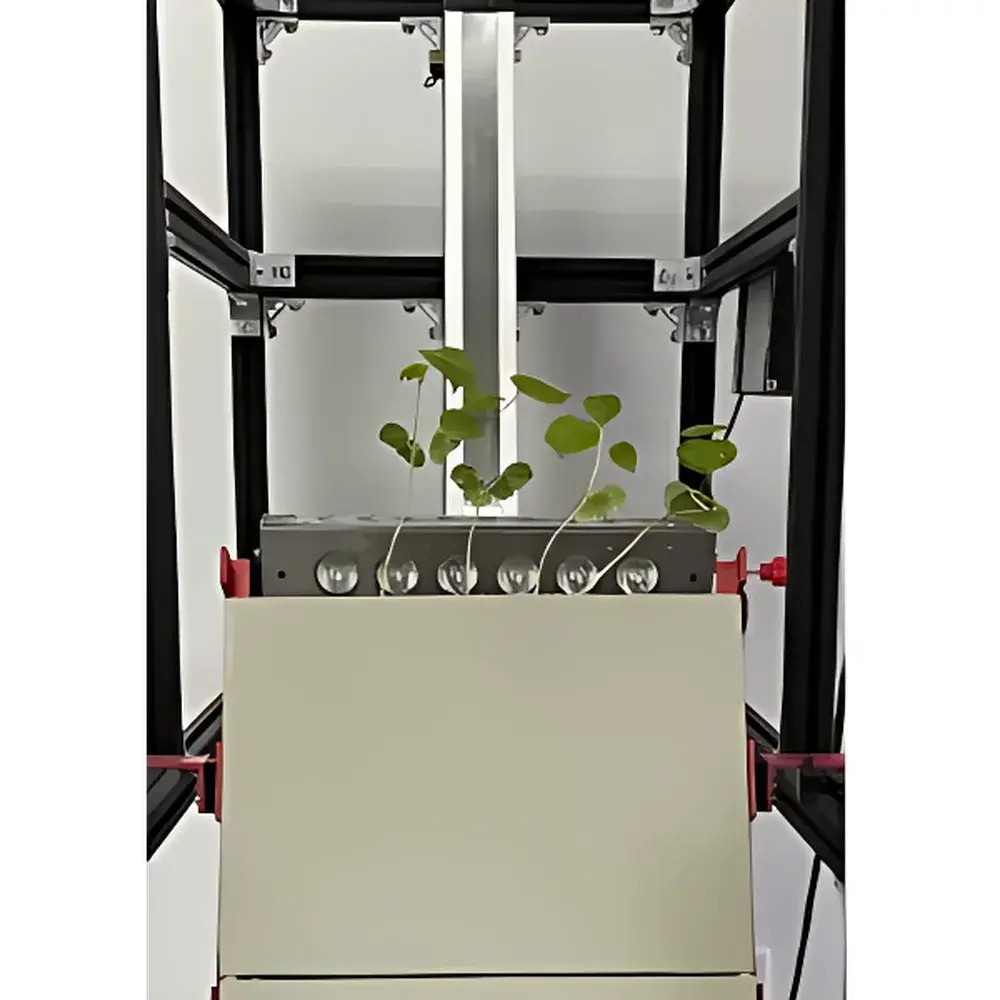
The EcoTech RhizoTron® Hyperspectral Imaging System is an engineered platform for non-invasive, in situ quantification of plant root architecture and rhizosphere biochemistry under controlled soil or semi-soil conditions. It integrates push-broom hyperspectral imaging (400–1000 nm standard, with optional 900–1700 nm SWIR extension) with synchronized UV-induced autofluorescence excitation (365 nm LED source) to resolve root structures that are spectrally indistinguishable from surrounding soil matrix using conventional RGB imaging. Unlike destructive excavation or limited-field-of-view minirhizotrons, the RhizoTron® operates on standardized RhizoBox® root observation chambers—transparent, soil-filled growth containers fitted with optical-grade viewing windows—enabling longitudinal, high-reproducibility monitoring of root development, senescence, pathogen colonization, and nutrient/water dynamics over weeks to months. Its measurement principle relies on spectral unmixing: differential absorption, scattering, and fluorescence emission profiles across hundreds of contiguous narrowband channels allow pixel-level discrimination of root tissue (e.g., cellulose, lignin, chlorophyll precursors, microbial metabolites) from mineral and organic soil components. This enables quantitative mapping of root length density, radial distribution, spectral indices (e.g., NDVI, NDSI, water index at 1450 nm), and biochemical proxies—including nitrogen status, moisture gradients, and decay-associated spectral shifts—without segmentation bias induced by low contrast.
Key Features
- Modular tilt-scanning platform with programmable motorized axis (1–40 mm/s speed, ±1 mm positional accuracy, 100 cm effective scan range); standard 60° angle aligns with natural root gravitropic orientation in RhizoBox® systems
- Dual-mode imaging capability: (a) Reflectance-based hyperspectral imaging (VIS-NIR: 400–1000 nm; SWIR: 900–1700 nm optional) and (b) UV-excited autofluorescence imaging (365 nm LED, bandpass-filtered detection) for enhanced root–soil contrast and metabolic activity assessment
- Integrated RGB imaging sensor (2448 × 2048 resolution) co-registered with hyperspectral data for multi-modal validation and morphological ground-truthing
- Expandable modular architecture: optional top-view modules include thermal-IR (640 × 512, −25°C to 150°C), chlorophyll fluorescence (720 × 560, 50 fps, full Kautsky protocol automation), and canopy-level hyperspectral/RGB imaging
- Intelligent LED cultivation stage (RGBW channels, 0–100% intensity control, up to 300 µmol/m²·s PPFD) supporting circadian rhythm simulation and spectral treatment experiments
- Embedded dual-control interface: touchscreen HMI + Windows-based GUI (full Chinese/English language support) with remote access via secure SSH or VPN-enabled API endpoints
Sample Compatibility & Compliance
The RhizoTron® is validated for use with standardized RhizoBox® root observation chambers (customizable dimensions: 10–60 cm height, 5–20 cm width), as well as user-fabricated acrylic or quartz-based root windows compatible with 0.5–2.0 mm optical thickness specifications. It accommodates both hydroponic agar/gel media and structured soil substrates (sand, loam, clay-loam mixtures up to 30% organic matter). All imaging protocols comply with OECD Test Guideline 208 (Terrestrial Plant Toxicity) and ISO 11269-2 for root growth assessment. Data acquisition workflows support audit trails required under FDA 21 CFR Part 11 (electronic records/signatures) when paired with SpectrAPP®’s optional compliance module. Raw spectral cubes (.bil/.hdr), processed morphology files (.csv/.json), and metadata logs are stored in FAIR-aligned directory structures compliant with MIAPPE v1.1 standards.
Software & Data Management
PhenoRoot™ software provides automated root morphology quantification—including total root length, convex hull area, root diameter distribution, cross-sectional density profiles, and branching topology—based on spectral-thresholded binary masks generated from hyperspectral indices. SpectrAPP® enables advanced spectral analytics: ROI-based spectral library matching, derivative spectroscopy (first/second order), spectral angle mapper (SAM), and custom index formulation (e.g., root decay index RDI = (R1649 − R1447) / (R1649 + R1447)). Both packages support batch processing, inter-session normalization, and export to Python/R environments via HDF5 or NetCDF4 formats. Optional integration with TensorFlow/PyTorch pipelines allows deployment of pre-trained deep learning models (e.g., U-Net for root segmentation, SAE for heavy metal prediction) directly within the acquisition workflow.
Applications
- Quantitative root phenotyping in breeding programs: genotype-by-environment interaction analysis for drought tolerance, phosphorus use efficiency, and root architecture QTL mapping
- Rhizosphere microbiome studies: spatial mapping of fluorescent pseudomonads (via UV autofluorescence) and fungal hyphae (using SWIR lignin/cellulose signatures)
- Soil–plant interaction modeling: high-resolution moisture distribution mapping (1450 nm water absorption band), nitrate/nitrite spectral deconvolution (800–900 nm), and organic carbon estimation
- Pathogen diagnostics: early detection of root rot progression (e.g., Aphanomyces euteiches, Fusarium solani) through time-series spectral shift analysis and machine-learned decay classification
- Heavy metal stress physiology: Pb/Cd/Cu concentration estimation in roots via SWIR-based regression models (R² > 0.92 in validation cohorts)
- Ecophysiological field validation: calibration transfer between controlled RhizoBox® data and minirhizotron or ground-penetrating radar field datasets
FAQ
What spectral ranges does the standard RhizoTron® system cover?
The base configuration includes a VIS-NIR hyperspectral camera covering 400–1000 nm at <5 nm spectral sampling interval. The 900–1700 nm SWIR extension is available as a factory-installed option.
Can the system operate without soil in the RhizoBox®?
Yes—it supports agar plates, filter paper, hydroponic gels, and aeroponic mist chambers. However, spectral contrast optimization and calibration protocols differ significantly from soil-based setups.
Is remote operation supported out of the box?
Remote control and scheduled unattended acquisition are enabled via Ethernet-connected embedded Linux controller. Full GUI mirroring requires optional VNC license and firewall configuration.
How is data traceability ensured for regulatory submissions?
SpectrAPP®’s optional 21 CFR Part 11 module logs all user actions, parameter changes, and processing steps with digital signatures and immutable timestamps—fully auditable for GLP/GMP submissions.
Does the system require darkroom conditions for UV imaging?
UV-induced autofluorescence imaging requires ambient light attenuation (<10 lux) during acquisition; integrated light-shielding curtains and timed LED pulsing minimize background interference.


