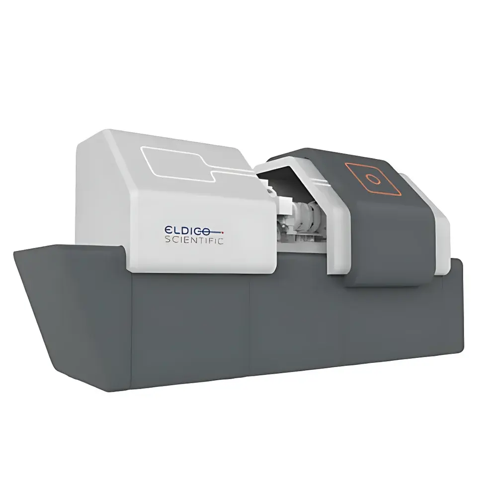
ELDICO ED-1 Three-Dimensional Microcrystal Electron Diffraction Instrument
| Brand | ELDICO Scientific AG |
|---|---|
| Origin | Switzerland |
| Model | ED-1 |
| Instrument Type | Single-Crystal Electron Diffractometer |
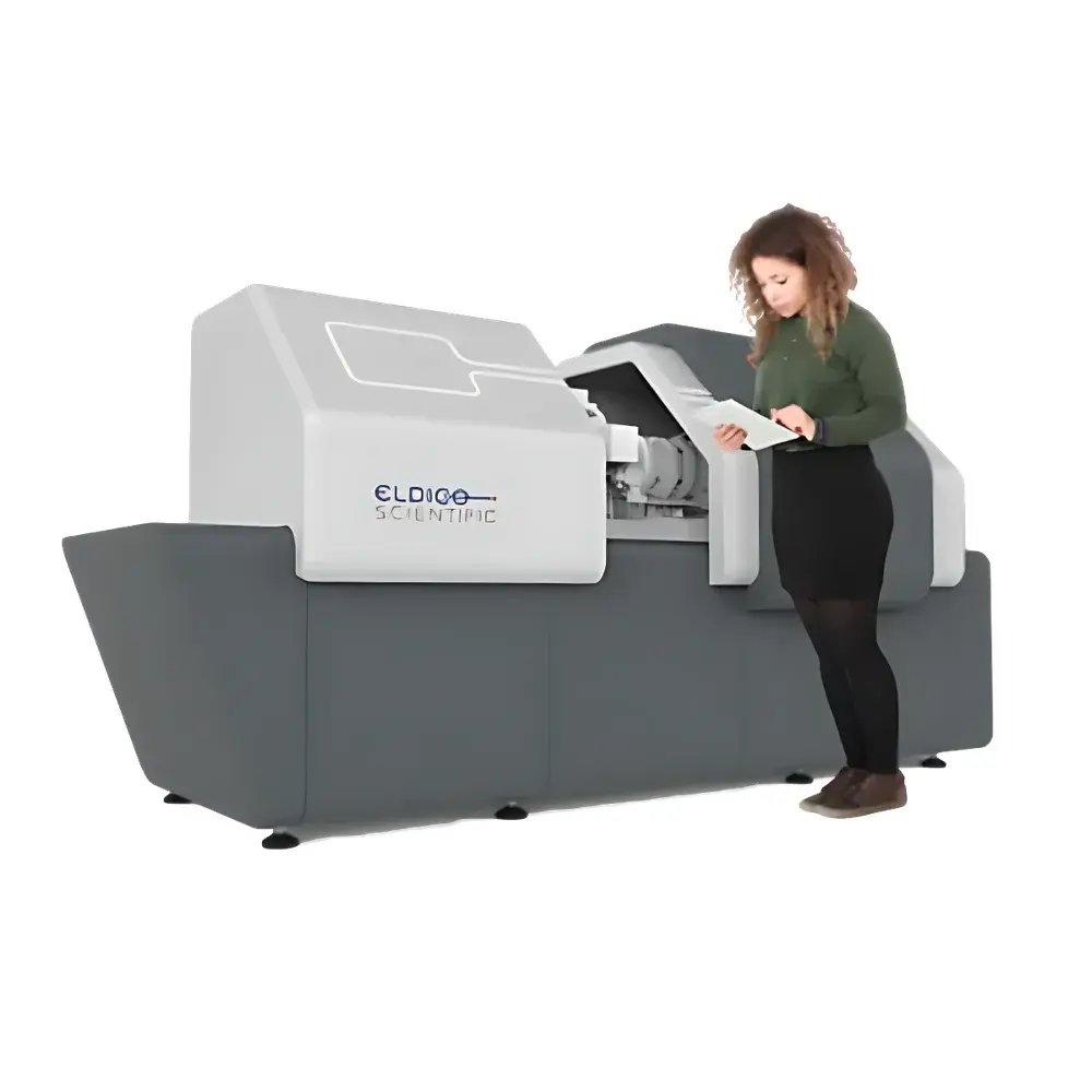
| Configuration | Floor-Standing |
| Resolution | 0.82 Å |
| Detector | DECTRIS QUADRO |
| Beam Energy | 160 keV |
| Sample Size Range | < 1000 nm |
Overview
The ELDICO ED-1 is a floor-standing, high-precision three-dimensional microcrystal electron diffraction (3D ED) instrument engineered for atomic-resolution structure determination of nanoscale crystalline materials. Unlike conventional single-crystal X-ray diffractometers—whose data collection requires crystals >10 µm in size—the ED-1 leverages 160 keV electrons to generate strong, coherent diffraction patterns from crystals as small as sub-100 nm. This capability stems from the ~10⁴ higher scattering cross-section of electrons compared to X-rays, enabling robust diffraction signal acquisition from vanishingly small, often polycrystalline or mosaic domains. The instrument implements continuous rotation electron diffraction (cRED) methodology, where a precisely controlled five-axis goniometer rotates the sample through 360° while collecting dynamic diffraction frames on a high-dynamic-range DECTRIS QUADRO hybrid pixel detector. Data are reconstructed into a full 3D reciprocal lattice, permitting ab initio structure solution and refinement using standard crystallographic software packages (e.g., SHELX, PHENIX, Olex2). Designed and manufactured by ELDICO Scientific AG—a Swiss company founded in 2019 with deep roots in electron crystallography—the ED-1 bridges a critical gap between transmission electron microscopy (TEM) and synchrotron-based diffraction, delivering laboratory-scale access to nanocrystallography without requiring cryo-TEM expertise or beamtime allocation.
Key Features
- Five-axis, sub-micron precision goniometer enabling full 360° continuous rotation for complete 3D reciprocal space sampling
- 160 keV high-brightness electron beam with optimized condenser optics for uniform illumination of nanoscale crystals
- Integrated DECTRIS QUADRO direct-detection electron camera offering high quantum efficiency (>80% at 160 keV), low noise, and frame rates up to 1 kHz
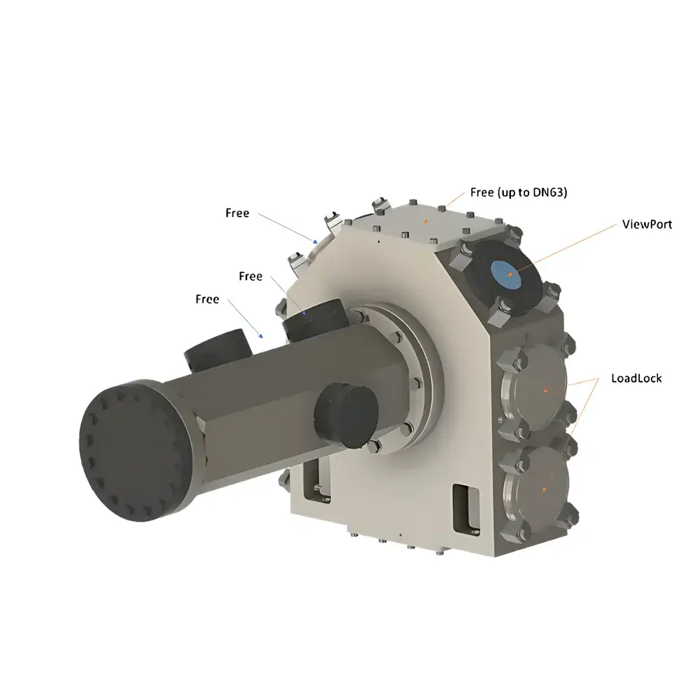
- Octagonal sample chamber architecture supporting modular expansion—including optional cryo-stages, in situ gas/liquid cells, and energy-dispersive X-ray spectroscopy (EDS) integration
- Automated sample loading and alignment routines reducing operator dependency and improving measurement reproducibility
- Real-time diffraction pattern preview and on-the-fly indexing via integrated ELDICO Control Suite software
Sample Compatibility & Compliance
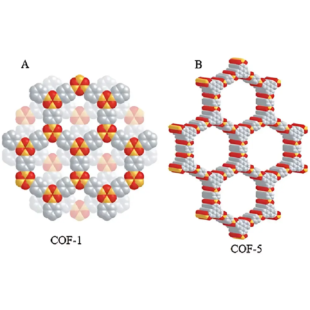
The ED-1 accommodates diverse crystalline specimens including metal–organic frameworks (MOFs), covalent organic frameworks (COFs), zeolites, pharmaceutical intermediates, chiral small molecules, and inorganic nanomaterials. It accepts standard TEM grids (e.g., Cu, Ni, Au), holey carbon films, and silicon nitride membranes. Sample stability under electron irradiation is managed via dose-fractionation protocols and beam blanking during stage movement—critical for radiation-sensitive organics. The system complies with IEC 61000-6-3 (EMC emission standards) and IEC 61000-6-4 (industrial immunity), and meets Swiss and EU CE requirements for laboratory electron optical equipment. While not classified as a medical device, its data output supports GLP-compliant structural reporting when used with audit-trail-enabled software configurations aligned with FDA 21 CFR Part 11 principles.
Software & Data Management
The ELDICO Control Suite provides unified control of beam parameters, goniometer motion, detector acquisition, and real-time data visualization. Raw .tiff or .h5 diffraction frames are exported in standardized formats compatible with CCP-EM, REDp, and PETS2 pipelines. Integrated metadata tagging includes timestamp, stage coordinates, exposure time, beam current, and detector gain settings—ensuring traceability for publication and regulatory submissions. Optional software modules support automated space-group determination, intensity integration (using XDS or DIALS), and seamless export to SHELXT/SHELXL or Olex2 for structure solution and refinement. All processing logs are archived with SHA-256 checksums, satisfying basic FAIR (Findable, Accessible, Interoperable, Reusable) data principles.
Applications
- De novo structure solution of nanocrystalline MOFs and COFs where bulk single crystals are inaccessible
- Absolute stereochemical assignment of chiral APIs and natural products via Flack parameter refinement
- Phase identification and quantification in multiphase crystalline mixtures (e.g., polymorphic blends, reaction intermediates)
- Crystallographic characterization of crystalline domains embedded in amorphous matrices or heterogeneous catalysts
- Time-resolved structural monitoring of solid-state reactions using in situ heating/cooling stages
- Validation of computational crystal structure predictions (CSP) against experimental 3D ED data
FAQ
What crystal size range is optimal for the ED-1?
Crystals between 50 nm and 800 nm yield highest-quality datasets; particles below 30 nm may require longer exposures or advanced denoising algorithms.
Can the ED-1 replace single-crystal X-ray diffraction (SC-XRD)?
It complements SC-XRD rather than replaces it: ED-1 excels where SC-XRD fails—nanoscale, twinned, or highly mosaic crystals—but SC-XRD remains preferred for high-accuracy bond-length/angle refinement of large, stable crystals.
Is cryogenic operation supported?
Yes—the octagonal chamber accepts commercially available cryo-transfer holders and Gatan 626/910 liquid-nitrogen stages for low-dose data collection at 100 K.
Does the system support automation for unattended data collection?
Fully supported via scriptable Python API and scheduled job queues; multi-sample carousels are available as factory-installed options.
How is radiation damage mitigated during data collection?
Through beam blanking during stage motion, dose-symmetric data collection strategies, and adaptive exposure control that dynamically adjusts frame time based on diffraction intensity decay profiles.


")