Evident SLIDEVIEW VS200 SILA Ultra-Fast Optical Sectioning Whole-Slide Imaging System
| Brand | Evident (formerly Olympus) |
|---|---|
| Origin | Japan |
| Manufacturer Type | Original Equipment Manufacturer (OEM) |
| Product Category | Imported Instrument |
| Model | SLIDEVIEW VS200 SILA |
| Instrument Type | Upright/Inverted Hybrid Microscope Platform |
Overview
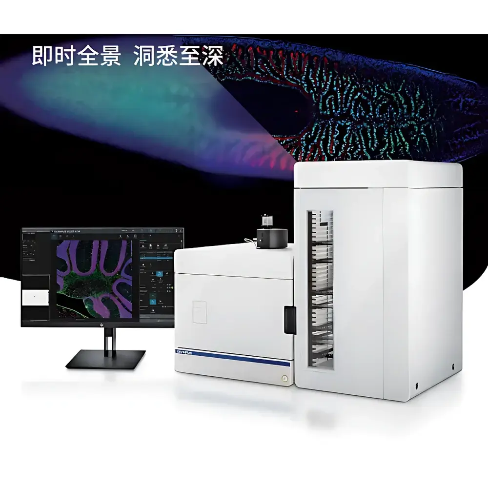
The Evident SLIDEVIEW VS200 SILA is an ultra-fast optical sectioning whole-slide imaging system engineered for high-fidelity, depth-resolved panoramic microscopy of thick biological specimens. Unlike conventional widefield scanners—whose out-of-focus blur limits contrast and structural clarity in specimens >20 µm—the VS200 SILA employs structured illumination with real-time computational optical sectioning to deliver near-confocal image quality without mechanical Z-scanning or post-acquisition deconvolution. Its core principle leverages laser-illuminated speckle-pattern projection combined with high-speed sCMOS acquisition and on-the-fly focal plane extraction algorithms. This enables true optical sectioning across large fields of view (up to 102 mm × 127 mm), preserving native signal-to-noise ratio while eliminating background haze. Designed specifically for neuroscience, developmental biology, oncology, and plant science applications, the system bridges the throughput–resolution gap between widefield slide scanners and point-scanning confocal microscopes—achieving up to 10× faster acquisition than conventional confocal platforms while maintaining subcellular detail in 3D-reconstructed volumes.
Key Features
- Optical sectioning capability with adjustable section thickness (5–50 µm), enabling crisp, haze-free imaging of thick sections (e.g., 40 µm mouse brain sagittal slices, organoids, plant tissue mounts)
- Hybrid upright/inverted microscope architecture supporting multiple contrast modalities: brightfield, fluorescence (widefield and optical-sectioned), darkfield, phase contrast, and simple polarized light
- High-throughput automated loader accommodating four standard slide formats: 26 × 76 mm, 52 × 76 mm, 76 × 102 mm, and 102 × 127 mm—enabling mixed-batch scanning of heterogeneous samples
- Real-time focal plane extraction using hardware-accelerated algorithms; no post-processing required for background suppression or Z-stack reconstruction
- Integrated TruAI deep learning module for user-trained region-of-interest (ROI) detection—including customizable CNN models for pancreatic islet identification, glomerular segmentation, or tumor margin delineation
- Maximum throughput of 210 slides per unattended run, with seamless integration into LIMS and digital pathology workflows
Sample Compatibility & Compliance
The VS200 SILA accommodates a broad spectrum of specimen types: FFPE and frozen tissue sections, whole-mount cleared tissues, cell monolayers, 3D organoid cultures, and botanical preparations. Its optical design ensures compatibility with standard fluorophores (DAPI, FITC, Cy3, TRITC, Alexa Fluor series) and label-free contrast mechanisms. The system conforms to ISO 13485:2016 for medical device quality management and supports audit trails compliant with FDA 21 CFR Part 11 when deployed in regulated environments. All firmware and software updates follow GLP/GMP-aligned validation protocols. While not a clinical diagnostic device per se, its imaging output meets the resolution and metadata standards required for research-grade digital pathology archives aligned with DICOM-SR and ASAM-Path guidelines.
Software & Data Management
Controlled via Evident’s VS-ASW (VS Advanced Software) platform, the system provides intuitive, parameter-minimized operation—requiring only one primary setting (optical section thickness) for routine use. Full Z-stack acquisition, maximum intensity projection (MIP), and pseudo-color depth encoding are performed in real time. Image data is exported in OME-TIFF format with embedded OMERO-compatible metadata, including objective magnification, illumination settings, exposure times, and spatial calibration. The TruAI module supports model import/export in ONNX format and includes built-in training workflows compatible with annotated datasets from QuPath or HALO. Data security features include role-based access control, encrypted local storage, and optional integration with enterprise PACS or cloud-based image repositories via DICOMweb or RESTful API.
Applications
The VS200 SILA serves as a foundational imaging platform across preclinical and translational research domains. In neuroscience, it enables rapid mapping of neuronal circuitry in serially sectioned brains with simultaneous multi-channel labeling (e.g., DAPI, GFAP, Iba1, NeuN). In cancer biology, it supports quantitative spatial analysis of tumor microenvironments—including immune cell infiltration patterns and stromal architecture—across entire tissue sections. Plant researchers utilize its large-field optical sectioning to visualize vascular bundles and meristematic zones in intact leaf or root mounts without physical sectioning. Developmental biologists apply it to time-series imaging of embryonic organogenesis in whole-mount specimens. Additionally, its compatibility with educational slide libraries and standardized histopathology training sets makes it suitable for academic teaching labs requiring reproducible, high-fidelity digital slide resources.
FAQ
How does VS200 SILA differ from traditional confocal slide scanners?
It eliminates point-scanning mechanics by using structured illumination and real-time computational sectioning—delivering comparable axial resolution at significantly higher speed and lower phototoxicity.
Can VS200 SILA perform true Z-stacks for 3D reconstruction?
Yes—it acquires full volumetric data sets with user-defined Z-intervals and exports native OME-TIFF stacks compatible with Imaris, Arivis Vision4D, and Fiji.
Is AI training supported for custom tissue annotations?
Yes—TruAI allows supervised training on user-provided ground-truth masks and deployment of inference models directly within the scanning workflow.
What slide formats does the automated loader support?
Four sizes: 26 × 76 mm, 52 × 76 mm, 76 × 102 mm, and 102 × 127 mm—all handled in a single unattended batch.
Does the system meet regulatory requirements for GxP environments?
While intended for research use, its software architecture supports 21 CFR Part 11 compliance through electronic signatures, audit trails, and change control documentation upon installation validation.
Related Products


