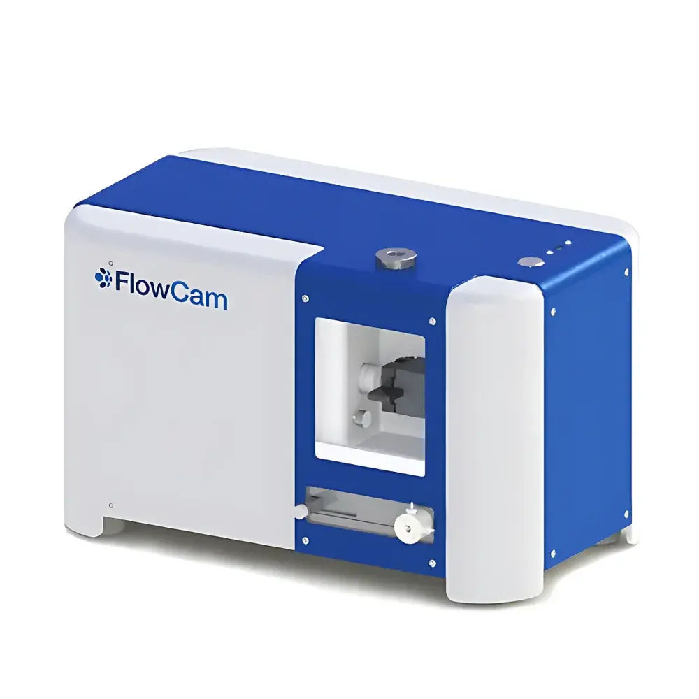
FlowCam 5000 Series Flow Imaging Microscope with Submicron-Capable Particle Analysis
| Brand | Yokogawa Fluid Imaging Technologies |
|---|---|
| Model | FlowCam 5000 |
| Detection Range | 3 µm – 300 µm |
| Optical Magnification Options | 4× (~40×), 10× (~100×), 20× (~200×) |
| Flow Cell Depth Options | 300 µm, 100 µm, 50 µm |
| Image Sensor | 1920 × 1200 pixel color CMOS |
| Frame Rate | Up to 100 fps |
| Minimum Detectable Feature Size | <1 µm (submicron-resolved morphology) |
| Sample Throughput | Up to 2 mL/min (at 4×) |
| Max Particle Concentration | 4 × 10⁶ particles/mL (for 3 µm particles) |
| Dimensions | 44 cm × 25 cm × 27 cm |
| Weight | 10 kg (operational), 24 kg (shipping) |
| Power Consumption | ≤92 W |
| Software | VisualSpreadsheet v6.x |
| Compliance | ALH-compatible, FDA 21 CFR Part 11 ready (audit trail & electronic signature optional), GLP/GMP-supportive workflow architecture |
Overview
The FlowCam 5000 Series is a compact, benchtop flow imaging microscope engineered for high-fidelity, real-time morphological characterization of suspended particles in liquid media. Unlike static image analysis or ensemble-averaging techniques (e.g., laser diffraction), the FlowCam 5000 employs hydrodynamic focusing and high-speed, wide-field optical imaging to capture individual particle images as they transit a precision-machined flow cell. This orthogonal approach—explicitly recommended by USP for subvisible particle analysis in biopharmaceuticals—enables direct measurement of size, shape, transparency, texture, and color at single-particle resolution. The system operates on the principle of flow cytometry combined with digital microscopy: particles are hydraulically aligned in laminar flow, illuminated via transmitted white light, and imaged across the full depth and width of the flow channel using a high-sensitivity color CMOS sensor. Each acquired image undergoes real-time segmentation, yielding quantifiable morphometric data without user intervention or thresholding bias.
Key Features
- Modular optical configuration: Select from 4×, 10×, or 20× objective lenses paired with matched flow cells (300 µm, 100 µm, or 50 µm depth) to optimize resolution and throughput for target particle size ranges (2–300 µm)
- Submicron-resolved morphology: Capable of resolving fine structural features below 1 µm—critical for distinguishing protein aggregates, silicone oil droplets, glass fragments, and microbial contaminants
- High-throughput imaging: Captures >10,000 particles per minute with automated focus stabilization and real-time image segmentation
- Integrated computing platform: Embedded industrial PC eliminates external hardware dependencies; boot-ready VisualSpreadsheet software launches directly from instrument firmware
- ALH (Automated Liquid Handling) compatibility: Supports integration with third-party autosamplers for unattended multi-sample workflows
- Color-accurate acquisition: 1920 × 1200 pixel RGB sensor with calibrated white-light illumination enables spectral ratio analysis (e.g., R/G, B/G, edge gradient) for material classification
- Ruggedized benchtop design: 10 kg operational weight, no vibration isolation required; validated for use in QC labs, field-deployable research vessels, and GMP manufacturing suites
Sample Compatibility & Compliance
The FlowCam 5000 accommodates aqueous and low-viscosity non-aqueous suspensions—including cell culture supernatants, buffer formulations, process streams, environmental water samples, and polymer dispersions—without requiring centrifugation, filtration, or staining. Its non-destructive, label-free methodology preserves sample integrity for downstream assays. The system supports regulatory compliance through configurable audit trails, user access controls, and electronic signature capability (when deployed with Part 11-compliant IT infrastructure). It meets analytical requirements outlined in USP , ISO 13322-2 (image analysis standards), and ASTM E2454 (microscopic particle counting). For biopharmaceutical applications, it provides orthogonal verification to light obscuration and membrane microscopy, fulfilling ICH Q5C stability assessment guidelines and supporting root-cause analysis of particulate-related batch failures.
Software & Data Management
VisualSpreadsheet v6.x serves as the unified interface for method development, acquisition control, image processing, and statistical reporting. It computes over 40 morphological descriptors—including area-equivalent diameter (ESD), aspect ratio, circularity (Hu moment-based), convexity, roughness, fiber straightness, and biovolume approximations—using ISO/IEC 17025-aligned algorithms. Users define custom classification rules via Boolean logic across parameter combinations (e.g., “circularity 50 µm² AND red/green ratio >1.8”) to isolate silicone oil, cellulose fibers, or diatom frustules. Batch comparison tools enable side-by-side histogram overlays, scatterplot clustering, and PCA-driven group separation. All raw images, metadata, and processed results export natively to CSV, TIFF, PDF, and HDF5 formats. Data integrity is maintained via immutable acquisition logs, timestamped versioning, and optional encrypted storage—fully compatible with LIMS and ELN integrations.
Applications
The FlowCam 5000 delivers actionable insights across regulated and research-intensive domains. In biopharmaceutical development, it characterizes protein aggregates, liposomal drug carriers, viral vectors, and host cell debris during upstream and downstream processing. In environmental science, it identifies and enumerates phytoplankton taxa (e.g., Microcystis, Anabaena, diatoms), microplastics, and fecal indicator organisms in raw and treated water. For materials science, it quantifies printer toner uniformity, CMP slurry abrasives, cement hydration products, and additive manufacturing powder sphericity. Food & beverage applications include yeast viability assessment, starch granule morphology, flavor encapsulant integrity, and contaminant screening (e.g., insect fragments, fungal hyphae). Marine ecologists rely on its validated taxonomy libraries for HAB monitoring, while QC labs deploy it for filter integrity testing, cleaning validation, and extractables/leachables profiling.
FAQ
What particle size range is resolvable with the FlowCam 5000?
The system detects particles from 3 µm to 300 µm in equivalent spherical diameter. With 20× optics and optimal contrast, submicron structural details (e.g., surface texture, internal granularity) are resolvable down to ~0.7 µm.
Can the FlowCam 5000 distinguish between silicone oil droplets and protein aggregates?
Yes—through combined analysis of transparency (optical density), edge gradient, circularity, and red/green intensity ratios, validated against reference standards per USP Annex A.
Is VisualSpreadsheet compliant with FDA 21 CFR Part 11?
The software supports Part 11 readiness when deployed on validated network infrastructure with role-based authentication, electronic signatures, and audit-trail logging enabled.
How does the FlowCam 5000 handle high-concentration samples?
A precision syringe pump maintains laminar flow velocity; maximum concentration limits (e.g., 4 × 10⁶ particles/mL at 3 µm) are defined by image overlap thresholds and are enforced in real time via software gating.
Does the system require daily calibration?
No routine calibration is required; NIST-traceable stage micrometers and certified particle standards (e.g., Duke Scientific) are used for periodic verification per ISO/IEC 17025.


