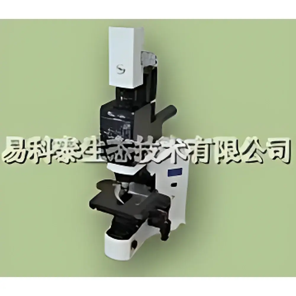
Fluorcam Microscopic Plant Chlorophyll Fluorescence Imaging System
| Brand | PSI (Czech Republic) |
|---|---|
| Origin | Czech Republic |
| Model | Fluorcam |
| Image Resolution | 12-bit, 512 × 512 pixels |
| Frame Rate | 50 fps |
| Data Interface | USB 2.0 |
| Spatial Resolution | ~1 mm (macro mode) |
| Excitation Sources | High-power LED arrays (450 nm, 625 nm, white), optional halogen lamp with shutter/LCD for continuous actinic light and saturating pulses |
| Detection | High-sensitivity cooled CCD camera with spectral response optimized for chlorophyll *a* fluorescence (680–750 nm) |
| Software | FluorCam 7.x with programmable protocol engine, ROI-based kinetic analysis, multi-parameter false-color mapping, and GLP-compliant audit trail (optional FDA 21 CFR Part 11 module) |
Overview
The Fluorcam Microscopic Plant Chlorophyll Fluorescence Imaging System is a high-resolution, quantitative imaging platform engineered for non-invasive, dynamic assessment of photosynthetic performance at cellular and subcellular scales. Based on pulse-amplitude modulated (PAM) fluorometry principles, the system captures spatially resolved chlorophyll *a* fluorescence kinetics—reflecting the functional status of Photosystem II (PSII)—under controlled photochemical and non-photochemical quenching conditions. Unlike conventional spot-measurement fluorometers, this system delivers two-dimensional fluorescence parameter maps (e.g., Fv/Fm, ΦPSII, NPQ, qP, qN, Rfd) across heterogeneous biological samples, enabling detection of localized stress responses, developmental gradients, or mutant phenotypes that remain invisible to bulk assays. Its modular optical architecture supports both macro-scale leaf-level imaging and microscopic integration—via coupling with upright or inverted research-grade microscopes—for subcellular resolution of chloroplasts, thylakoid stacks, or even single grana.
Key Features
- High-speed acquisition: 50 frames per second (fps) at full 512 × 512 resolution, enabling capture of rapid transient events including QA− reoxidation kinetics and antenna connectivity dynamics.
- Multi-wavelength excitation: Configurable LED panels (450 nm blue, 625 nm red, broad-spectrum white) allow selective excitation of specific light-harvesting complexes (LHCII, CP24, CP26) to dissect contributions to photochemical vs. non-photochemical quenching.
- Integrated actinic & saturating light control: Halogen-based continuous illumination system with precision electronic shutter or LCD-based pulse modulation ensures reproducible induction of photosynthetic electron transport and PSII closure.
- Subcellular sensitivity: Cooled, low-noise CCD sensor with quantum efficiency >75% in the 680–750 nm emission band enables low-light (<1 µmol photons·m−2·s−1) imaging—minimizing perturbation of native metabolic states while resolving fluorescence from individual chloroplasts or stromal compartments.
- Modular microscope compatibility: Designed for seamless integration with standard epifluorescence, confocal, or light-sheet microscopes; includes mechanical adapters, synchronization TTL triggers, and optical path calibration routines.
Sample Compatibility & Compliance
The Fluorcam Microscopic System accommodates diverse biological specimens—from isolated protoplasts and algal colonies to intact leaves, seedlings, and tissue sections—without requiring exogenous dyes or genetic modification. Its open-stage design permits use with Petri dishes, glass-bottom culture plates, or custom sample holders. All hardware and firmware comply with CE, RoHS, and IEC 61000-6-3 electromagnetic compatibility standards. The FluorCam 7.x software optionally supports 21 CFR Part 11 compliance (electronic signatures, audit trail, user access control) for regulated environments including GLP and GMP plant biotechnology labs. Data export formats include TIFF (16-bit), HDF5, and CSV—ensuring interoperability with MATLAB, Python (NumPy/SciPy), and FIJI/ImageJ workflows.
Software & Data Management
FluorCam 7.x provides a fully scriptable, experiment-driven interface built on a real-time acquisition kernel. Users define complex protocols—including variable light intensities, dark adaptation sequences, multiple saturating pulses, and time-lapse intervals—via intuitive drag-and-drop modules or Python-based scripting. The software performs automated ROI segmentation, kinetic curve extraction per pixel or region, and synchronized parametric mapping across all acquired frames. Built-in algorithms compute over 20 standardized fluorescence parameters (including F0, Fm, Fv, F0′, Fm′, ΦPSII, NPQ, qP, qN, Rfd) with pixel-wise statistical confidence reporting. Raw data are stored with embedded metadata (timestamp, light settings, camera gain, temperature), and version-controlled analysis pipelines ensure full traceability from raw image to publication-ready figure.
Applications
- Early detection of abiotic stress (drought, heat, heavy metals, UV-B) via spatial heterogeneity in NPQ distribution and PSII quantum yield.
- Functional phenotyping of Arabidopsis, rice, or crop mutants—mapping chloroplast-level defects in electron transport or photoprotective capacity.
- Quantitative analysis of chloroplast movement, division, and retrograde signaling under varying light regimes.
- Validation of transgenic lines expressing fluorescent protein reporters (e.g., GFP-tagged PSII subunits) using dual-channel excitation/emission separation.
- Time-resolved studies of state transitions (qT), energy spillover between PSII and PSI, and cyclic electron flow activation kinetics.
FAQ
Can the system image non-chlorophyll fluorescence, such as GFP or synthetic dyes?
Yes—the excitation LED array and emission filter wheel support customizable wavelength configurations; users may install bandpass filters (e.g., 480/30 nm excitation + 535/40 nm emission) for GFP, YFP, or DAPI imaging without hardware modification.
Is microscope integration plug-and-play or does it require custom alignment?
Integration requires optical path calibration (provided in the manual) and mechanical adapter installation; PSI supplies alignment targets and step-by-step SOPs—no third-party optics tuning is needed.
What is the minimum detectable fluorescence change (ΔF/F0) under low-light imaging conditions?
System noise floor corresponds to <0.5% ΔF/F0 at 10-ms integration time, validated using calibrated neutral density filters and reference fluorophore solutions.
Does the software support batch processing of time-series datasets from multiple genotypes or treatments?
Yes—FluorCam 7.x includes a batch analysis wizard that applies identical ROI definitions, parameter calculations, and normalization routines across hundreds of image stacks, generating consolidated Excel reports with ANOVA-ready statistics.
Are calibration certificates and IQ/OQ documentation available for regulated laboratories?
Factory calibration certificates (intensity, timing, spatial uniformity) are provided with each system; IQ/OQ protocols and validation templates compliant with ISO/IEC 17025 are available upon request for qualified users.


