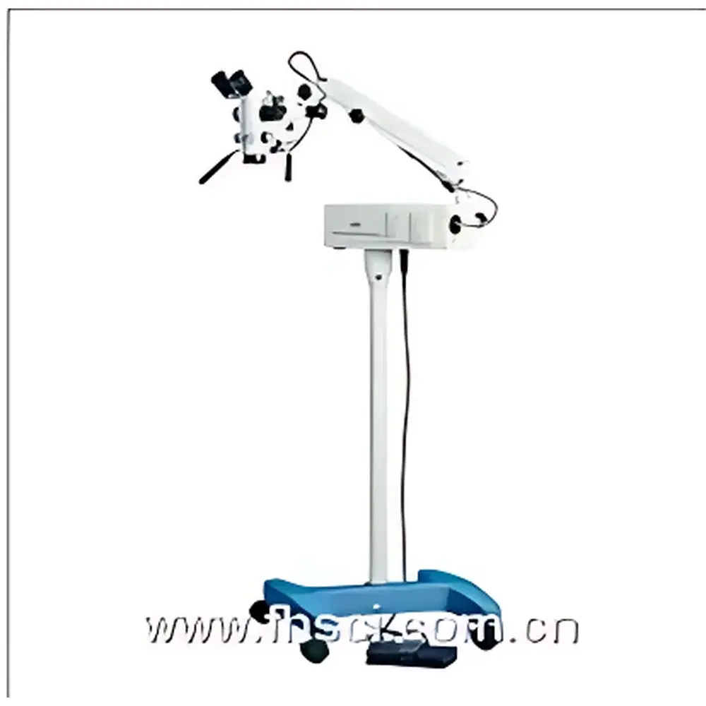
Harvard Apparatus PSMB5 / PSMT5 Precision Surgical Microscope
| Brand | Harvard Apparatus |
|---|---|
| Origin | USA |
| Model | PSMB5 / PSMT5 |
| Total Optical Magnification | 1.8×–40× |
| Eyepiece | 12.5× |
| Interpupillary Distance Adjustment | 50–70 mm |
| Diopter Adjustment | ±6 D |
| Working Distance (Focus Range) | 94–344 mm |
| Objective Focal Length | 200 mm (PSMB5), 350 mm (PSMT5, recommended for 103 cm mounting height) |
| Field of View | 58–10 mm (5-step zoom) |
| Fine Focus Travel | 30 mm |
| Articulated Arm Reach | Max. 870 mm radius |
| Vertical Travel Range | 700–1100 mm |
| Mounting Height Options | 89 cm or 103 cm (user-specified at order) |
| Dual Halogen Illumination | 12 V, 100 W cold mirror lamps with fiber-optic light guides |
| Camera Interface | C-mount optical port compatible with 1/2″ CCD sensors (e.g., WPI COLCAM, DC2000M) |
| Power Input | 110 V or 220 V, 50–60 Hz |
| Net Weight | 43 kg (94 lb) |
Overview
The Harvard Apparatus PSMB5 and PSMT5 are precision-engineered stereoscopic surgical microscopes designed for high-fidelity visualization in preclinical neuroscience, microvascular surgery, and small-animal experimental procedures. Built upon a robust yet lightweight articulated arm architecture, these instruments employ coaxial Galilean optical design with five discrete magnification steps (3.2×, 5×, 8×, 13×, 20×) and continuous fine-tuning via motorized foot-controlled focus—functionality typically reserved for premium-tier clinical systems. The optical path delivers diffraction-limited resolution across the full magnification range, with high-contrast image formation enabled by apochromatic correction, anti-reflective coated lenses, and optimized light transmission through cold-mirror dichroic beam splitters. Both models support dual independent halogen illumination sources (12 V, 100 W), each coupled to the objective via flexible fiber-optic light guides—ensuring stable, shadow-free, color-accurate illumination without thermal load on the specimen. The PSMB5 is configured with a 200 mm focal-length objective for standard working distances (89 cm mounting), while the PSMT5 integrates a 350 mm objective optimized for extended vertical reach (103 cm mounting), accommodating deeper anatomical access in rodent or larger murine models.
Key Features
- Motorized foot-switch focus control for hands-free depth adjustment during delicate manipulations
- Five-step optical zoom system (3.2×–20×) with fixed magnification positions and smooth interpolation between steps
- Adjustable interpupillary distance (50–70 mm) and ±6 diopter eyepiece compensation for operator-specific visual acuity calibration
- Dual independent halogen lamp modules with cold-mirror reflectors and rapid, power-intact source switching—eliminating illumination downtime during procedure transitions
- C-mount optical port supporting 1/2″ format digital cameras (e.g., WPI COLCAM, DC2000M) for real-time monitoring, documentation, and synchronized video recording
- Compact, low-inertia mechanical arm with 870 mm maximum horizontal reach and 400 mm vertical travel (700–1100 mm range), enabling precise repositioning without destabilizing the surgical field
- Modular mounting options: standardized 89 cm or 103 cm base height configurations—selected at time of order to match experimental setup ergonomics and objective focal length requirements
Sample Compatibility & Compliance
The PSMB5/PSMT5 platform is validated for use in GLP-compliant preclinical research environments and aligns with core optical safety and performance expectations outlined in ISO 10940:2018 (Surgical microscopes — Requirements and test methods) and ANSI Z80.10-2020 (Ophthalmic instruments — Surgical microscopes). Its non-contact visualization geometry, adjustable working distance (94–344 mm), and wide field-of-view progression (58 mm down to 10 mm) accommodate diverse specimen sizes—from neonatal mouse craniotomies to adult rat spinal cord exposure. All optical components comply with RoHS Directive 2011/65/EU and meet IEC 60601-1 third edition electrical safety standards. The dual-illumination architecture satisfies IEC 62471 photobiological safety classification for Group 1 (exempt) risk level under normal operating conditions.
Software & Data Management
While the microscope operates as a standalone optical instrument, its integrated C-mount interface enables seamless integration with third-party acquisition software platforms—including NIS-Elements (Nikon), MetaMorph (Molecular Devices), and WPI’s own AcqKnowledge-compatible drivers. When paired with compliant digital cameras, the system supports timestamped image capture, frame-locked video export (AVI, TIFF stack), and metadata embedding (magnification, illumination intensity, focus position). Audit trails for camera settings and user-defined presets can be maintained in accordance with FDA 21 CFR Part 11 requirements when deployed within validated laboratory information management systems (LIMS).
Applications
- Microdissection and intracranial injection in transgenic mouse models
- Carotid artery ligation, femoral vein cannulation, and microvascular anastomosis in rats
- Retinal surgery simulation and optic nerve crush modeling
- Electrophysiology probe placement under visual guidance (e.g., silicon probe insertion, microwire electrode targeting)
- Live-tissue fluorescence-assisted surgery using external excitation sources coupled via side-port adapters
- Training modules for neurosurgical technique development in academic and contract research organizations (CROs)
FAQ
What is the difference between PSMB5 and PSMT5?
The PSMB5 uses a 200 mm focal-length objective and is intended for installation at the 89 cm mounting height. The PSMT5 features a 350 mm objective and is engineered for the 103 cm mounting configuration, providing greater working distance and improved depth-of-field for deeper surgical access.
Is motorized focus programmable or recallable?
No—the foot-switch focus system is analog and manually actuated; it does not store positions or interface with external controllers. For automated focus sequencing, integration with third-party motorized stage controllers is required.
Can this microscope be used with fluorescence imaging?
Yes—via optional side-port excitation couplers and filter cubes. The base optical path is broadband-transmissive (350–1000 nm), and the cold-mirror illumination system minimizes UV/IR heat load on specimens during prolonged exposure.
Does the system include camera and recording software?
No—the microscope ships with the C-mount adapter only. Cameras and acquisition software must be selected and purchased separately based on resolution, frame rate, and compatibility requirements.
What maintenance is required for long-term optical stability?
Annual verification of collimation and magnification calibration is recommended. Optical surfaces should be cleaned using lens-grade solvents and lint-free wipes; halogen lamps should be replaced in matched pairs to maintain balanced illumination intensity and color temperature.


