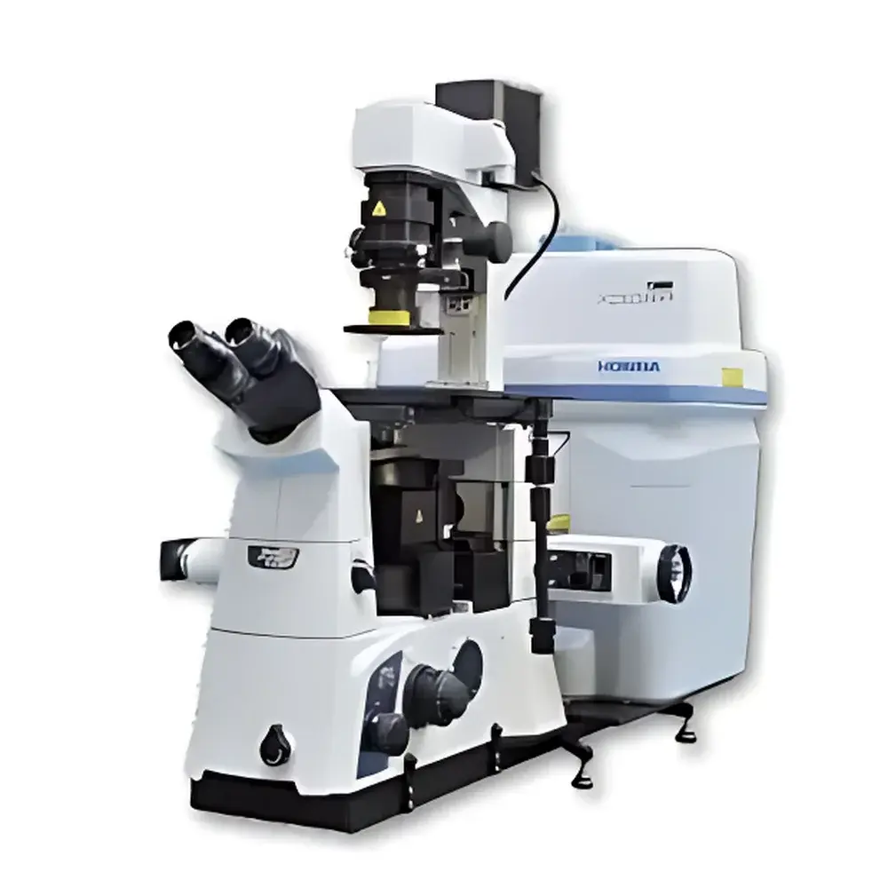
HORIBA LabRAM XploRA INV Inverted Confocal Raman Microscope
| Brand | HORIBA |
|---|---|
| Origin | France |
| Model | LabRAM XploRA INV |
| Instrument Type | Inverted Confocal Raman Microscope |
| Laser Options | 532 nm, 638 nm, 785 nm (multi-laser auto-switching) |
| Grating Options | 4 gratings auto-switched (600, 1200, 1800, 2400 gr/mm) |
| Spectral Range | 250–10,000 cm⁻¹ (configurable per laser/grating combination) |
| Spatial Resolution | Sub-micron (diffraction-limited, dependent on objective and wavelength) |
| Automation | Auto-focus, auto-exposure, auto-calibration, self-diagnostic |
| Software Compliance | FDA 21 CFR Part 11-ready audit trail, GLP/GMP-supportive workflow logging |
| Optional Modules | DuoScan™ high-speed Raman imaging, SWIFT™ fast spectral acquisition, epifluorescence integration, polarization control, precision XYZ stage, environmental stages (cryo/heated/biological incubation), AFM-Raman coupling interface |
Overview
The HORIBA LabRAM XploRA INV is an inverted confocal Raman microscope engineered for high-fidelity molecular characterization of delicate, hydrated, or live biological specimens under near-native conditions. Unlike conventional upright configurations, its inverted optical architecture provides unobstructed access to the sample plane—enabling seamless integration with micromanipulators, optical tweezers, microfluidic chambers, and cell culture hardware. The system operates on the principle of inelastic light scattering (Raman effect), where monochromatic laser excitation induces vibrational mode shifts in molecular bonds, generating a fingerprint-like spectrum that reveals chemical composition, crystallinity, stress/strain states, and molecular conformation. Its confocal design delivers axial optical sectioning with rejection of out-of-focus signal, ensuring depth-resolved spectral acquisition at micron-scale spatial resolution. Designed for laboratories requiring rigorous reproducibility and regulatory traceability, the XploRA INV meets foundational requirements for GLP-compliant research and preclinical assay development.
Key Features
- Research-grade inverted microscope platform with high-NA objectives optimized for aqueous immersion and low-fluorescence glass-bottom dishes.
- Triple-laser module with fully automated switching between 532 nm, 638 nm, and 785 nm excitation—minimizing photodamage while maximizing signal-to-noise across diverse sample types (e.g., 785 nm for reduced fluorescence in tissues; 532 nm for high-resolution inorganic analysis).
- Four-grating turret with motorized selection (600, 1200, 1800, 2400 grooves/mm), enabling flexible trade-offs between spectral range (up to 10,000 cm⁻¹), resolution (<1 cm⁻¹ FWHM with high-density gratings), and throughput.
- Integrated DuoScan™ technology: a galvanometric mirror-based scanning system delivering diffraction-limited Raman imaging at speeds exceeding 100 spectra/sec without compromising spatial fidelity—ideal for dynamic cellular processes or large-area tissue mapping.
- SWIFT™ (Sequential Wave Imaging Fourier Transform) mode for rapid hyperspectral acquisition with real-time spectral reconstruction and background subtraction.
- Full automation suite: GO! software assistant initiates end-to-end workflows—including auto-alignment, laser power optimization, focus stabilization via motorized Z-stage feedback, exposure time adaptation, and daily calibration verification against NIST-traceable standards.
Sample Compatibility & Compliance
The XploRA INV accommodates live cells in CO₂-controlled incubators, 3D organoids in Matrigel, microtissues on flexible substrates, and subcellular organelles within intact neurons—all without sample dehydration or metal coating. Its open-access stage supports custom-built accessories: patch-clamp rigs, temperature-controlled chambers (−196 °C to +600 °C), humidity-regulated enclosures, and piezoelectric nanopositioners. Regulatory readiness includes built-in audit trail generation, electronic signature support, and user-access-level permissions aligned with FDA 21 CFR Part 11 Annex 11 expectations. All calibration routines are documented with timestamps, operator IDs, and deviation logs—fully compatible with ISO/IEC 17025 quality management systems and pharmaceutical QC environments.
Software & Data Management
LabSpec 6 software provides a unified interface for acquisition, multivariate analysis (PCA, MCR-ALS), spectral library matching (with >20,000 reference compounds), and correlative imaging (Raman + epifluorescence overlay). Raw data is stored in vendor-neutral HDF5 format with embedded metadata (laser power, grating ID, objective magnification, environmental parameters). Batch processing scripts support automated QA/QC checks—e.g., peak intensity stability across time-series experiments or wavenumber drift correction using internal silicon reference. Export modules comply with ASTM E131 and ISO 18385 spectral data interchange standards.
Applications
- Label-free identification of intracellular lipid droplets, amyloid aggregates, and drug metabolites in live primary neurons.
- Mapping chemical heterogeneity in tumor spheroids—distinguishing necrotic core, proliferative rim, and hypoxic gradients via CH/CC band ratios.
- In situ monitoring of polymer degradation kinetics under thermal cycling or UV exposure.
- Strain distribution analysis in 2D materials (graphene, MoS₂) transferred onto flexible substrates.
- Quantitative assessment of calcification in vascular smooth muscle cells under osteogenic induction.
- Correlative Raman–fluorescence imaging of mitochondrial redox state (NADH/FAD⁺ ratio) alongside pH-sensitive dyes.
FAQ
Can the XploRA INV be used for time-lapse Raman imaging of live cells?
Yes—the system supports continuous acquisition with adaptive exposure control and active focus stabilization, enabling hour-long kinetic studies with minimal phototoxicity.
Is AFM-Raman coupling supported natively?
The platform features standardized mechanical and electrical interfaces for third-party AFM integration; HORIBA-certified alignment protocols and synchronized trigger I/O ensure co-registered topographic–chemical datasets.
Does the software support multivariate statistical modeling for classification tasks?
LabSpec 6 includes PCA, hierarchical clustering, and supervised PLS-DA modules with cross-validation tools—validated against USP guidelines for chemometric model validation.
How is spectral calibration traceability maintained?
Daily auto-calibration uses a fused silica reference and Si wafer peak (520.7 cm⁻¹); all calibrations are logged with NIST-traceable uncertainty budgets and archived with raw spectra.
Can environmental stages be controlled directly from LabSpec?
Yes—via native drivers for Linkam, Instec, and Quantum Design stages, including real-time temperature ramping profiles and closed-loop feedback during acquisition.