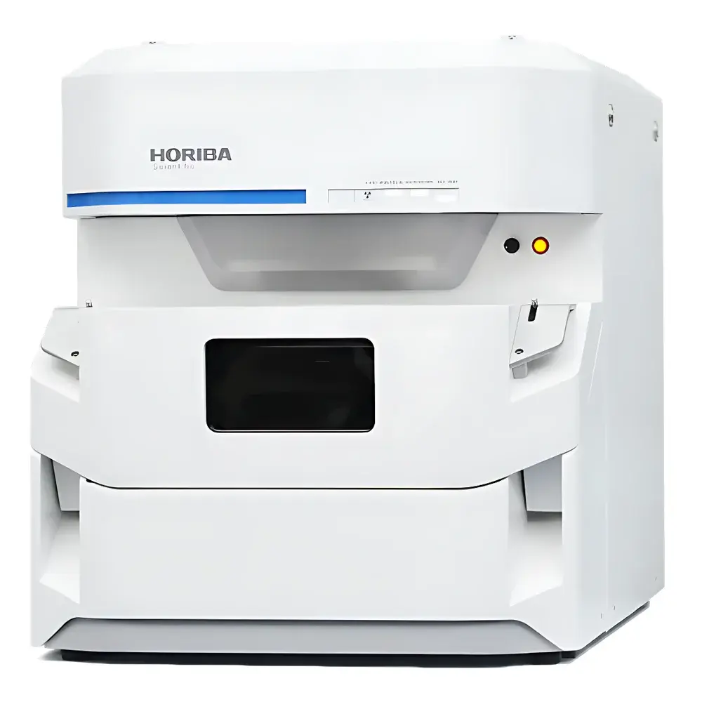
HORIBA XGT-9000 Wavelength Dispersive X-Ray Fluorescence Microscope
| Brand | HORIBA |
|---|---|
| Origin | Japan |
| Manufacturer Type | Original Equipment Manufacturer (OEM) |
| Product Origin | Imported |
| Model | XGT-9000 |
| Excitation Mode | Single-Wavelength Excitation |
| X-ray Tube Power | 50 W |
| Voltage Range | Up to 50 kV |
| Current Range | Up to 1000 µA |
| Elemental Coverage | Na (11) to U (92) |
| Minimum Spot Size | ≥10 µm |
| Optional Capillary Probes | 15 µm and 100 µm |
| Detector Configuration | Dual-mode — Energy-Dispersive XRF + Transmission X-ray Imaging |
| Sample Chamber Flexibility | Up to 4 configurable measurement atmospheres (air, He, vacuum, N₂) |
| Software Platform | HORIBA X-RAY LAB |
Overview
The HORIBA XGT-9000 is a high-performance wavelength dispersive X-ray fluorescence (WDXRF) microscope engineered for non-destructive, spatially resolved elemental analysis at the microscale. Unlike conventional benchtop XRF systems, the XGT-9000 integrates a focused microbeam excitation source with dual-detection capability—simultaneously acquiring fluorescent X-ray spectra for elemental mapping and transmission X-ray images for structural morphology. Its core architecture leverages a high-brightness microfocus X-ray tube (50 kV / 1000 µA), coupled with precision-polished capillary optics that deliver spot sizes down to 10 µm—enabling quantitative microanalysis of heterogeneous materials without sectioning or coating. The instrument operates on fundamental principles of characteristic X-ray emission following inner-shell ionization, with spectral resolution optimized for peak separation across the full Na–U range. Designed for laboratories requiring trace-level sensitivity in complex matrices, the XGT-9000 supports both qualitative identification and semi-quantitative mapping under standardized conditions compliant with ISO 17025-accredited workflows.
Key Features
- Microbeam excitation system with selectable spot sizes from 10 µm to 1.2 mm—enabling seamless transition between micro-area analysis and macro-scale survey scanning
- Dual-mode detection: integrated energy-dispersive XRF detector for elemental distribution imaging + transmission X-ray detector for internal structure visualization
- High-intensity capillary optics options: 15 µm probe for high-spatial-resolution mapping; 100 µm probe for rapid large-area screening
- Multi-atmosphere sample chamber supporting air, helium purge, nitrogen, and high-vacuum environments—critical for light-element (Na–F) detection and low-background measurements
- High-resolution optical imaging subsystem with digital zoom, multi-illumination modes (brightfield, darkfield, oblique), and real-time overlay of XRF maps onto optical coordinates
- Rugged mechanical stage with motorized XYZ positioning and sub-micron repeatability for automated raster scanning and multi-point analysis
Sample Compatibility & Compliance
The XGT-9000 accommodates diverse physical forms—including solid fragments, PCBs, coins, wafers, thin films, powders, liquids (in sealed cells), and biological specimens—without requirement for conductive coating or vacuum compatibility constraints. Optional sample holders include tilt stages, rotating mounts, and custom fixtures for irregular geometries. All measurement protocols are fully traceable and support audit-ready documentation per GLP and GMP requirements. Data acquisition complies with ISO 21043 (XRF spectrometry), ASTM E1621 (standard guide for XRF analysis), and USP / for elemental impurities in pharmaceuticals. When configured with optional RoHS screening module, analyses meet EU Directive 2011/65/EU compliance thresholds for Pb, Cd, Hg, Cr(VI), and Br-containing flame retardants.
Software & Data Management
HORIBA X-RAY LAB software provides an integrated environment for instrument control, spectral processing, elemental mapping, and report generation. The interface supports customizable multi-monitor layouts, synchronized display of optical images, spectra, periodic table overlays, and false-color elemental maps. All raw spectra and metadata are stored in vendor-neutral HDF5 format, ensuring long-term archival integrity. Built-in calibration tools enable matrix-matched standardization using certified reference materials (CRMs), while advanced deconvolution algorithms correct for inter-element absorption and enhancement effects. Optional modules extend functionality: multilayer film thickness analysis (for coatings and semiconductor stacks), automated batch queue execution (for unattended overnight runs), particle recognition and co-localization analysis (for inclusion studies), and LabSpec 6 export for principal component analysis (PCA) and cluster identification. Audit trail logging satisfies FDA 21 CFR Part 11 requirements for electronic records and signatures.
Applications
- Failure analysis of electronic components: correlating solder joint voids (via transmission mode) with localized Cu/Sn/Pb segregation (via XRF mapping)
- Geological and gemological research: identifying trace-element zoning in mineral inclusions and distinguishing synthetic vs. natural gemstones
- Materials science: quantifying dopant distributions in battery cathodes, catalyst nanoparticles, and thin-film photovoltaics
- Forensic science: micro-spatial profiling of gunshot residue particles, paint chips, and document inks
- Environmental monitoring: detecting heavy-metal contamination in soil particulates and airborne filters at sub-ppm levels
- Pharmaceutical quality control: verifying elemental impurity profiles in active pharmaceutical ingredients (APIs) per ICH Q3D guidelines
FAQ
What is the smallest detectable feature size achievable with the XGT-9000?
The system achieves a minimum excitation spot size of 10 µm using standard capillary optics; optional high-intensity probes offer 15 µm resolution with enhanced photon flux for faster dwell times.
Can the XGT-9000 quantify light elements such as sodium or oxygen?
Yes—when operated under helium purge or vacuum atmosphere, the instrument reliably detects and maps elements from Na (Z=11) through U (Z=92), with optimized spectral deconvolution for overlapping L/M lines.
Is the transmission X-ray imaging mode capable of tomographic reconstruction?
No—the current configuration acquires single-angle projection images only; however, the mechanical stage supports user-defined angular increments for custom CT workflows via external scripting.
How does the XGT-9000 handle sample charging during analysis of insulating materials?
The instrument employs low-energy electron flood gun compensation and adjustable beam current settings to minimize surface charge accumulation without requiring carbon coating.
Does HORIBA provide application-specific method development support?
Yes—HORIBA’s Applications Lab offers remote and on-site method validation services, including CRM selection, matrix-matched calibration, limit-of-detection (LOD) determination, and SOP documentation aligned with ISO/IEC 17025 accreditation requirements.
Related Products